Article Text

Abstract
This study reports on a series of patients who were diagnosed as having had a transient lateral patellar dislocation by magnetic resonance imaging (MRI). The images were reviewed with specific reference to the medial collateral ligament (MCL), a heretofore undescribed concomitant injury. Eighty patients were diagnosed on MRI as having had transient lateral patellar dislocation. Their mean age was 23.9 years (SD 7.5). Forty patients (50.0%) had co-existent MCL injuries. These injuries were classified as grade 1 (n = 20), grade 2 (n = 17) and grade 3 (n = 3). These results suggest that MCL injury commonly accompanies transient lateral patella dislocation, most likely due to a shared valgus injury. It appears to occur more commonly in male patients and if unidentified may explain both delayed recovery and persistent morbidity in more severe cases. In this setting, without specifically excluding co-existent MCL injury, the current vogue for early rehabilitation should be adopted with caution.
Statistics from Altmetric.com
Patellar dislocation accounts for 2–3% of all knee injuries1 and is seen as the second most common cause of knee haemarthrosis.2 Despite this, its pathophysiology is poorly understood and its management continues to remain controversial. The main ongoing problem is recurrence but it is still far from certain which if any population is most at risk for this and if surgery diminishes this risk.3
Traditionally, this is an injury that has been associated with women although this has been challenged in some reports.4 5 This association is felt to be due to a relatively increased genu valgum seen in women6 7 although it is unclear if any particular sport represents a specific risk.8 Furthermore, it has been described mainly with patients in their second decade of life,5 9 although Atkin et al5 do stress the potential bias in such demographic statements. It is possible that decreasing neuromuscular control over the lower limb10 11 may contribute to a patient's susceptibility but it would appear difficult to provide age-specific aetiologies. However, it is also an injury that can be transient in nature, with patients unaware it has happened. Indeed, magnetic resonance imaging (MRI) can often offer the first diagnosis with its characteristic pattern of lateral femoral condyle bone bruising.12
The established risk factors for patellar dislocation are many and well documented. These include trochlear dysplasia,13 medial patellofemoral ligament insufficiency,14 patella alta,15 abnormal Q angle,16 genu valgum,17 generalised ligamentous laxity18 and iliotibial band dysfunction.19 A relationship between patellar dislocation and patellar maltracking is also quoted, and this is felt to be due to an imbalance between the vastus medialis and vastus lateralis.20 21 Unequal forces across the knee have also led to eccentric histological changes seen in patients with chronic patellar tendinitis22 and this may also be an association with patellar dislocation.
Treatment of this condition can be simplified into conservative and surgical modalities. Redislocation rates with these methods have been quoted at 0–47% and 0–32%, respectively.1 There are currently over 100 documented surgical procedures described, and apart from recurrence complications have only been associated with operative interventions.23 In relation to conservative management, the emphasis is placed on aggressive, early mobilisation of the patient.24
Stefancin and Parker1 produced a meta-analysis of the 70 published papers on this subject in 2007. The authors noted that due to the multifactorial element of the condition and debate about management, multiple confounders existed in many studies. However, in an attempt to summarise treatment, they advocated conservative management, except in cases when associated with osteochondral fractures, substantial disruption of medial patellar stabilisers, lateral subluxation of the patella compared with the contralateral knee, second dislocation or failure to respond to rehabilitation.1
The medial patellar stabilisers are both static, such as the inherent bony anatomy25 and dynamic, namely the medial retinaculum,26 medial patellofemoral ligament (which accounts for 60% of medial stability)27 and the vastus medialis obliquus (VMO).28 However, to date, the authors are unaware of any studies looking at the medial collateral ligament (MCL) and its association with this injury complex in patellar dislocation.
The aim of this MRI-based study was to establish an incidence of MCL injury in patients with transient patellar dislocation and to examine if there was a significant age or gender association.
Materials and methods
During the study period from November 2001 to April 2008 inclusive, 80 patients were diagnosed with transient patellar dislocation, confirmed by MRI scanning. In all cases, the radiological diagnosis correlated with the clinical findings in these patients. No patient had any previous diagnoses of patellar dislocation.
All patients were imaged in a Philips new generation Gyroscan Intera Tesla (T) scanner (Koninklijke Philips Electronics Ltd, Eindhoven, The Netherlands) and had coronal short T1 inversion recovery (STIR), coronal T1 and sagittal and axial T2 images performed.
The resultant scans were reported on by a musculoskeletal fellowship-trained radiologist. The radiological diagnosis of transient patellar dislocation was confirmed by the presence of the characteristic countercoup pattern of lateral femoral condyle bone bruising (fig 1). Following this, the status of the other structures in the knee was commented upon.
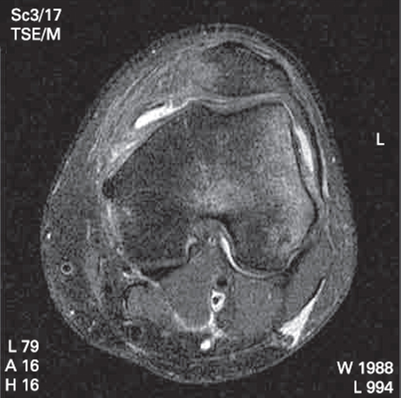
Axial T2 image of the knee showing lateral femoral condyle and medial patellar facet bone bruising consistent with a diagnosis of transient patellar dislocation.
In particular, the status of the MCL was noted. Injuries to this structure were classified as grades 1-3 as per the classification of Schweitzer et al.29 This system defines grade 1 injuries as having fluid on the inner or outer aspect of the superficial fibres. Grade 2 injuries show fluid on both sides of the ligament with or without internal foci of high signal but without discontinuity, whereas grade 3 injuries are classified as having complete disruption/discontinuity of the ligament (table 1).
MRI findings seen in injuries of the MCL
Statistical analysis was performed initially between the no-injury and the injury group using a two-sample t-test. Then, looking specifically at the injury group, a linear model was fitted to age with covariates gender, injury group and the interaction gender by injury group. The first term assesses the effect of gender on age, the second the effect of injury group on age and the last term allows for a different effect of injury group between men and women. In an alternative analysis, as the number with grade 3 MCL injuries was so small, the Wilcoxon rank sum test was used in comparisons involving this group; p values of p<0.05 were regarded as statistically significant. All analyses were carried out using SAS version 9.1.3.
Results
Eighty patients imaged during the study period had a radiological diagnosis of transient patellar dislocation. This group consisted of 66 men and 14 women. The mean age of the total study cohort was 23.9 years (SD 7.5, range 11–60). The mean ages of the male group was 23.7 years (SD 5.9, range 14–41) and of the female group was 25.0 years (SD 12.9, range 11–60).
All 80 patients had bone bruising of their lateral femoral condyle in a pattern consistent with transient patellar dislocation. In addition, patellar derangements were described in 40 of the patients. These took the form of bone bruising to the medial facet (n = 30) and chondral injuries (n = 10). The chondral injuries were subdivided according to their location and consisted of the junction of the medial and lateral facets (n = 3), the lateral facet (n = 3), the medial facet (n = 2). In the other two cases, medial facet bone oedema was observed in association with a chondral injury to the junction of the medial and lateral facet (n = 1) and to the medial facet (n = 1).
Of specific relevance to this study was the MCL and any attendant damage to it. This is an association that has not previously been described to the authors' best knowledge. The MCL was found to be injured in 40 cases. The mean age of patients with MCL injuries was 23.5 years (SD 5.1, range 15–36). There were 36 men with a mean age of 23.8 years (SD 5.1, range 15–36) and four women with a mean age of 20.8 years (SD 4.6, range 15–25).
Looking at the groups overall, the non-MCL injury group had a mean age of 24.3 years (SD 9.4, range 11–60). This compares with a mean age of 23.5 years (SD 5.1, range 15–36) for the MCL injury group. When these groups were compared, no significant differences were found among men (p = 0.85), women (p = 0.46) or overall (p = 0.63). Moreover, there was no significant difference between the male no-injury and female no-injury groups (p = 0.33).
Out of the 40 patients with MCL damage, 20 were classified as grade 1 (fig 2). The mean age of this group was 21.6 years (SD 5.1, range 15–36). There were 17 male patients with a mean age of 21.7 years (SD 5.3, range 15–36). The remaining three female patients had a mean age of 19.3 years (SD 4.5, range 15–24).

Coronal short T1 inversion recovery image showing a grade 1 medial collateral ligament injury as characterised by fluid on the inner side only of the superficial fibres of the ligament.
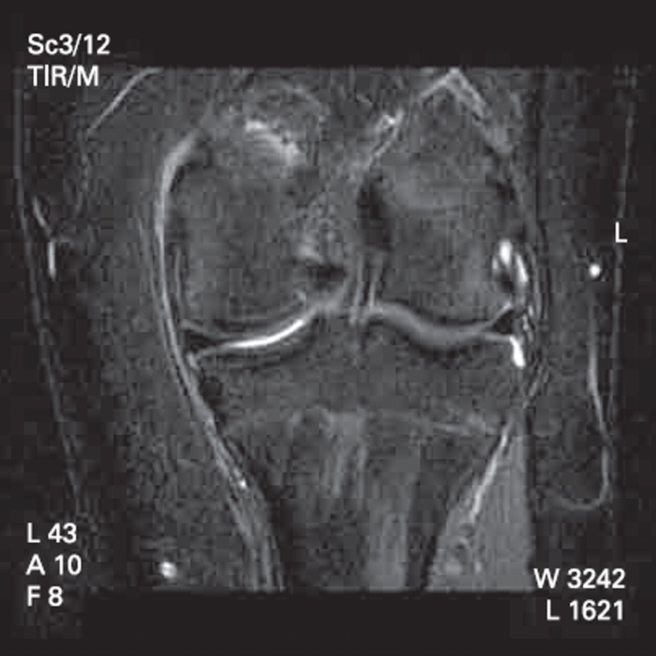
Grade 2 MCL injuries (fig 3) were described in 17 patients. This group had a mean age of 25.9 years (SD 4.3, range 16–32). Sixteen of these patients were men with a mean age of 26.0 years (SD 4.4, range 16–32). The sole female patient with a grade 2 injury was 25 years of age. There were three male patients in the grade 3 subgroup (fig 4). Their mean age was 24.0 years (SD 2, range 22–26).

Coronal short T1 inversion recovery image showing a grade 2 medial collateral ligament injury as characterised by fluid on both sides of the superficial fibres of the ligament. The ligament remains in continuity.

Coronal short T1 inversion recovery image showing a grade 3 medial collateral ligament injury as characterised by fluid on both sides of the ligament associated with a discontinuity of the ligament.
When the MCL injury group was analysed, a linear model was fitted to age with covariates gender, injury group and gender by injury group. The interaction term gender by injury group was not significant, ie, the age differences between injury groups was the same for men and women (p = 0.81). The effect of gender was not significant (p = 0.43), ie, there was no significant difference in age between men and women. There was a significant difference between injury groups with patients, with grade 1 MCL injuries having a significantly lower age than those in the grade 2 group (p = 0.005), and when adjusted for multiple comparisons (three) (p = 0.015). The difference was 4.59 years, with a standard error of 1.54. In an alternative analysis, as the number with grade 3 MCL damage was so small the Wilcoxon rank sum test was used to compare separately patients in the grade 1 and grade 3 groups and the p value was p = 0.17. Comparing the grade 2 and grade 3 groups the difference was also not significant (p = 0.33).
Discussion
The mean age of 23.9 years (SD 7.5) in this study as well as the subgroup mean ages of 24.3 years (SD 9.4) in the non-MCL injury group and 23.5 years (SD 5.1) in the patients with MCL injuries is older than the traditionally described second decade.5 9 It is also older than the mean 21.5 years stated in the review by Stefancin and Parker.1 Furthermore, the preponderance of 66 men overall and 36 men in the MCL injury group is also in contrast to the majority of the studies on the subject.1
Whereas the association between patellar dislocations and MCL injuries has not to the authors' knowledge previously been described, Hunter et al30 published a series of surgically repaired MCL injuries and looked at the incidence of vastus medialis injury. They reported on a consecutive series of 189 moderate and severe MCL injuries that underwent open repair. In their series, they noted a 21% (40 patients) incidence of VMO disruption. They concluded that a significant relationship exists between acute MCL injury and damage to the VMO muscle. However, no specific correlation is drawn in relation to the reverse of this relationship.
The details of patients' sports were not recorded in this study, although there were no professional sportspeople in the series. However, the three highest participation sports in the reporting country are Gaelic games, soccer and rugby union. Studies of injuries in these sports31,–,33 show that the lower limb accounts for 52.7–87% of all injuries. In particular, the knee is involved in 12–17% of injuries. Soft tissue injuries account for 55–81% of injuries in total. In relation to rugby union injuries,32 39% of knee injuries were ligamentous and 75% of these involved the MCL. It would appear reasonable to presume that this study cohort follows similar patterns.
Due to the retrospective nature of this study, it was not possible to collate data in relation to the contralateral limb or in relation to limb dominance. However, it is worth noting that many players are equally adept at kicking with both feet, with Cromwell et al31 citing 34% of players who kicked with both feet in Gaelic football. This may make limb dominance a moot issue.
It is also worth considering the effect of training and conditioning on the support structures of the knee. Exercise has been seen to increase the mechanical and structural properties of ligaments.34 However, overuse can have a detrimental effect on injury status, as seen in other anatomical areas such as the groin35 and other sports such as Australian Rules football.36 In essence, the cohort in this study is a self-selecting one in that patients with clearly defined patellar pathology have been removed before scanning. The inherent difficulties in specifying exact mechanisms of injury and individual sporting details in groups such as this have been alluded to by others.31 32
This study presents a series of injured MCL based on MRI findings. Although all patients underwent clinical examination of their knee before scanning, they were not specifically assessed for this injury. However, many authors have described a high correlation between MRI and clinical findings of MCL damage37,–,40 while accepting that MRI images may suggest a higher grade of injury than is clinically apparent.41
What this study adds
MCL injury does occur with transient patellar dislocation. It should be examined for, especially in male patients, and treatment of the dislocation may need to be amended if it is found to be present.
The majority of MCL injuries heal with conservative management and lead to no adverse sequelae for the patient.42 43 However, a small number can go on to cause chronic medial laxity of the knee and require surgical intervention.44 Without recognition of the injury, it is difficult to predict which knees will become chronically lax.
In summary, the results of this paper present strong evidence for the existence of a relationship between patellar dislocation and MCL injury. It is to be presumed that while the MCL plays no role in maintaining patellar stability, its role as the primary restraint to valgus in the knee45 puts it at risk during the mechanism of injury of patellar dislocation. In particular, when the demographics of the patients in this study are analysed, an MCL injury should be especially suspected in male patients and patients who might be considered older than the standard for patellar dislocation. In these patients, who may be considered outside the normal demographics for patellar dislocation, aggressive rehabilitation of the patellar dislocation injury should be treated with at least circumspection and should perhaps be avoided.
References
Footnotes
-
Competing interests None.