Article Text

Abstract
Introduction Tissue barriers function as “gate keepers” between different compartments (usually blood and tissue) and are created by specialised membrane-associated proteins, located at the lateral plasma membrane of epithelial and endothelial cells. By sealing the paracellular space, such barriers impede the free diffusion of solutes and molecules across epithelial and endothelial monolayers, thereby creating an organ-specific homeostatic milieu.
Tendon cells originate from yet poorly described precursor cells and develop in a particular “niche” close to vascular walls. Degenerative processes, trauma and injury severely disturb the internal milieu of the niche. Concomitantly, pathological alterations including hyperproliferation, erroneous tendon cell differentiation, and calcification take place [Aström, 1995; Oliva, 2012].
It is a commonly held belief that the vasculature plays a crucial role in creating and maintaining a favourable milieu for tendon cells to develop, however, experimental evidence of how this is accomplished has been lacking.
We now report that tendon cells are protected from the systemic circulation by a novel barrier, the blood-tendon barrier (BTB), located in the vascular wall of tendon vessels. The diffusion restraint exerted by the BTB is capable of defining and controlling the molecular composition of the tendon-specific niche, which after all governs proper tendon cell differentiation [Bi, 2007].
Methods RT-PCR, immunohistochemistry, transmission electron microscopy and in vivo tracer perfusion were performed to identify the expression of barrier-associated proteins and to demonstrate the “tightness” of tendon vessels in human Palmaris longus and mouse Achilles tendons.

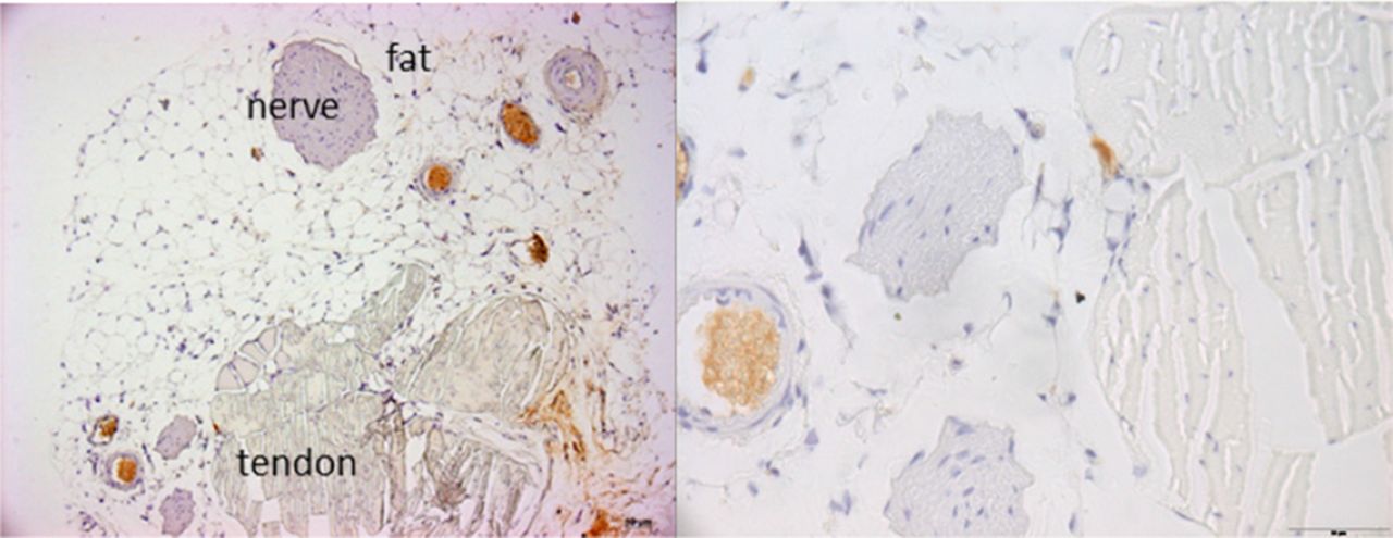
Murine Achilles tendon blood vessels are tight to a 10kD biotinylated dextran tracer
Results Perfusion with defined tracer substances revealed that the blood-tendon-barrier impedes the passive transport of macromolecules (≥ 10 kDa) from the blood stream to the surrounding tendon tissue (Figure 1), but is permeable to molecules <287D. The expression of barrier-related proteins, such as zonula occludens protein-1 (ZO-1), occludin, claudin-3, -5, and-12, in human and murine tendon vascular cells further corroborates the assumption that a restrictive tissue barrier acts at the blood-tendon interface.
Discussion The mechanisms and factors maintaining the internal milieu of the developmental niche in healthy tendons are not yet fully understood. Here, we describe a novel vascular structure, the blood-tendon barrer (BTB), which separates tendon tissue from the systemic circulation in intact human and murine tendons. The role of this structure in tendinopathy, tendon regeneration and tendon development remains to be elucidated.
References Aström, et al. Clin Orthop Relat Res. 1995;316:151–164, 1995
Bi, et al. Nat Med. 2007;13(10):1219–1227
Oliva, et al. BMC Med. 2012;10:95