Article Text

Abstract
Introduction We have previously shown that human and rat tendon cells produce insulin and secrete it upon glucose stimulation. Moreover, the level of tendon insulin production is affected by the amount of glucose taken up by nutrition [Lehner, 2012]. We now hypothesise that nutritional glucose affects tendon healing in a rat model.
Methods In 60 female Lewis rats full thickness defects were created in one Achilles tendon and left unsutured.
The rats were randomly assigned to three groups, one was fed a high glucose diet, one a diet with low glucose/high fat and one a control diet, for 2 weeks each. Before surgery, one and two weeks after, gait analysis was performed using a NoldusTM catwalk system. After two weeks the animals were sacrificed and tendon size was measured and tendons were biomechanically tested an evaluated by various histological methods.
Results Gait Analysis revealed a significant difference between the three groups one week after surgery. The intermediate toe spread (the range between second and fourth toe, a measure for the load on the limb) of the high glucose group is significantly increased one week p.o. (0,49 cm ± 0,07; n = 20; p < 0.01) compared to the control group (0,42 cm ± 0,08; n = 19) and to the high fat group (0,40 cm ± 0,12; n = 20) (Figure 1).
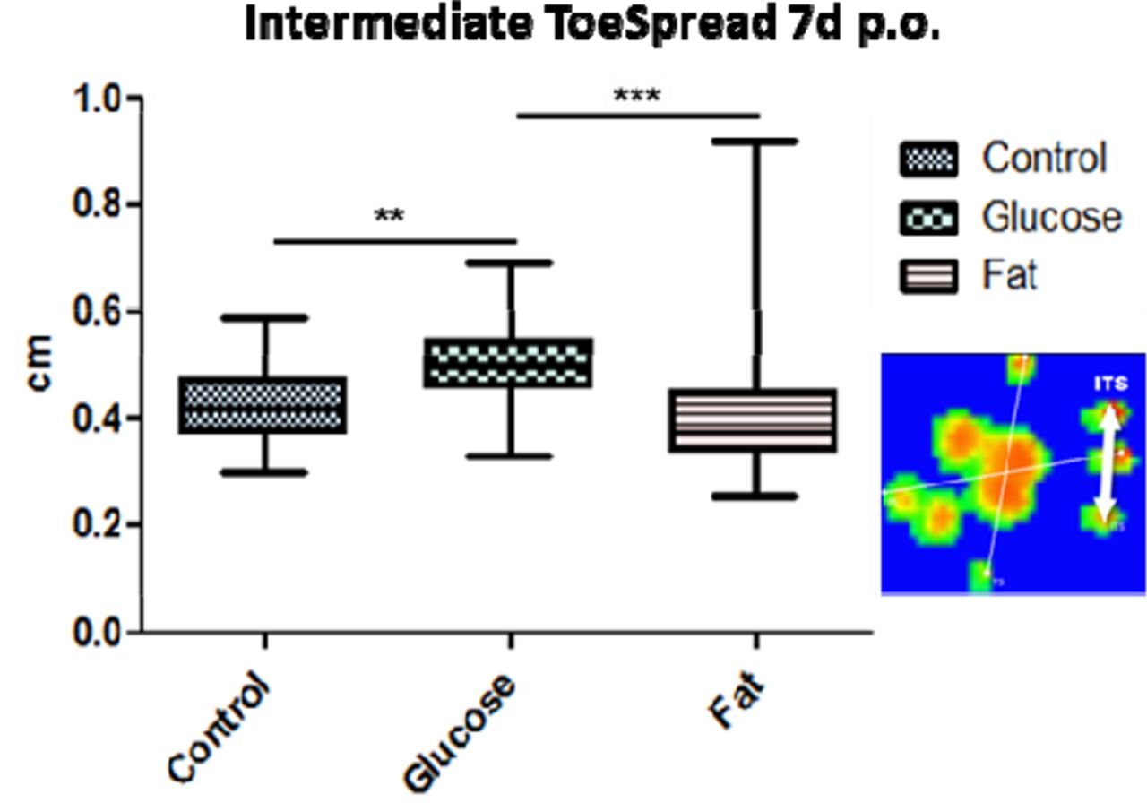
The intermediate toe spread (ITS) of the injured limb is significantly increased in the high glucose group
Measurement of length and thickness of the newly formed tissue revealed a significant (p < 0.001) difference in tendon thickness of the newly formed tissue between the high-glucose group (4,26 mm ± 0,29; n = 20) and the control group (3,66 mm ± 0,39; n = 19) as well as between the high glucose and the high-fat group (4,32 mm ± 0,20; n = 20).
Biomechanical testing revealed no significant difference between the groups in maximum tensile load, however, the new fibrous tissue from the glucose group is significantly (p < 0.05) stiffer (20,82 N/mm ± 8.08; n = 14) compared to the control group (15,07 N/mm ± 4.32; n = 14) (Figure 2). The stiffness of these tendons was similar to the stiffness of intact tendon tissue of the control group (20,63 N/mm ± 10,96; n = 14).

The regenerated fibrous tissue in the rats fed with high glucose diet is significantly (p < 0.05) stiffer compared to the control and the high fat group
Discussion Newly formed tendon tissue quality is affected by nutritional glucose. This finding is relevant for understanding diabetes related tendinopathy. Nutritional parameters may account for the interindividual variation of tendon quality and regeneration. The underlying molecular mechanisms will be examined.
Reference Lehner et al. Horm Metab Res. 2012;44:506–510