Article Text

Abstract
Introduction Tendon has a very slow self-healing capacity after injury 1 and tendon-derived stem cells (TDSCs) can be used in tendon regeneration and tendon wound healing.2 In in-vivo situation and during inflammation, tendon could be subjected to oxidative stress. Studies show that oxidative stress can cause apoptosis in tendon fibroblast and contribute to tendon degeneration.3 Also, oxidative stress can cause senescence in mesenchymal stem cells.4 However, the effects of oxidative stress on tendon-derived stem cells are still unknown. The aim of this study is to investigate the effect of oxidative stress (hydrogen peroxide) on proliferation and differentiation of TDSCs.
Methods Proliferation Assays
TDSCs were cultured in expansion medium with 0, 0.1 and 0.5 mM hydrogen peroxide (H2O2) with a density of 100 cells/ cm.2 The total number of cells in each well was counted by haemocytometer every two days.
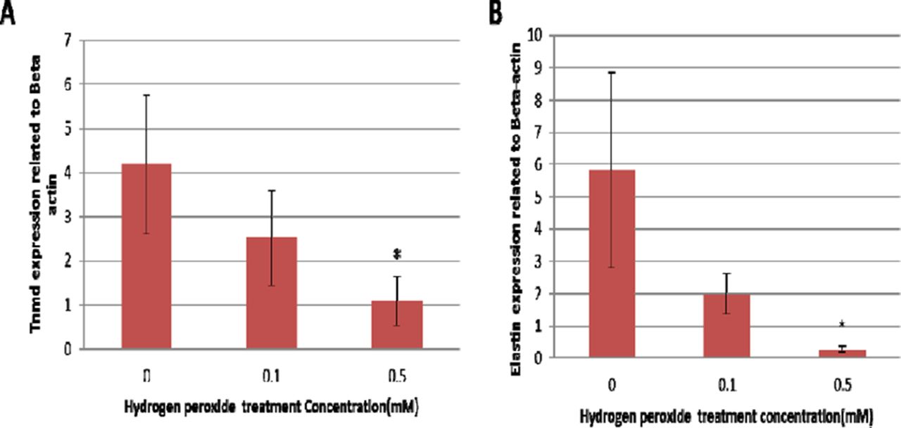
Tenogenic differentiation in oxidative environment TDSCs were cultured in expansion medium until confluence and then the medium changed to tenogenic medium (expansion medium plus 25ng/ml cTGF and 25 mM Ascorbic acid) to induce tenogenic differentiation for 14 days with or without the presence of H2O2 (0.1 or 0.5 mM). TDSCs cultured in expansion medium served as a control. Four tenogenic markers, Tenomodulin (Tnmd), Elastin (Eln), Scleraxis (Scx) and Collagen type one (Col1a1) were examined by real time PCR
Alizarin red S staining To test if oxidative stress will drive TDSCs towards osteogenic differentiation in tenogenic niche. 2% Alizarin Red S solution was used to indicate the formation of calcium nodules in TDSCs cultured 21 days in tenogenic medium.
Results and discussion The proliferation rate of TDSCs is significantly lower in 0.5 mM H2O2 treatment while the difference is not statistically significant in 0.1 mM treatment compared with 0 mM group (Figure 1).

Graph for the proliferation rate of TDSCs
Tenogenic differentiation The expression for Tnmd and Eln is significantly decreased (Figure 2) while no significant difference for the expression of Col1a1 and Scx (Data not shown). This implies that high oxidative stress may have adverse effect on TDSC differentiation.
(A) Tnmd expression. (B) Eln expression
Alizarin Red S Staining Small amount of Calcium nodules formed in all the four groups and the absorbance for 0.5 mM H2O2 treatment group is statistically significant compared with 0 mM H2O2 group (data not shown). This implies that oxidative stress may drive TDSCs towards osteogenesis.
Acknowledgement This research was made possible by resources donated by Lui Che Woo Foundation Limited.
References 1 Brandl et al. J Exp Cell Res 2011;317:1541–1547
2 Ni et al. J Biomaterials 2013;(34):2024–2037
3 Sharama et al. J Musculoskelet Neuronal Interact 2006;6(2):181–190
4 Yuan et al. BBA 2003;1641:35–41