Article Text

Abstract
Introduction One of the causes of anterior groin pain is known to be related to insertional tensor fascia lata (TFL) tendinopathy (soft tissue origin). This entity has been very well described a few years ago in ultrasound (US) examination by Bass and Connell as an insertional tendinopathy in the upper lateral iliac crest, demonstrating the ultrasound findings (US) produced in such a pathology. In this report we present two clinical cases that came to our office consulting for chronic groin pain as a new entity in TFL tendinopathy.
Method The two cases were active males (30 and 39 years old). Clinical symptoms were longstanding pain and inability for running. MRI studies were done and none of them described any affection of tensor fascia lata tendon (neither of the studies focused in specific TFL tendon). One of the patient (30 yrs.) was diagnosed as having femoroacetabular impingement and underwent surgery for this hip before came to us demonstrating a very unsatisfactory outcome.
Specific Sonograms for TFL were obtained with the patient lying in a decubitus position with the symptomatic side uppermost. After confirming the main area of pain as the proximal anterolateral area of thigh, we did and US examination with a Sonosite M-Turbo® HFL-50 (6–15 MHz) linear transducer. The US exam area began in the anterior iliac crest and scans were obtained in both longitudinal and transverse planes.
Results The proximal “insertional” TFL tendon presented normal size and echogenicity (compared contralateral) in both cases.
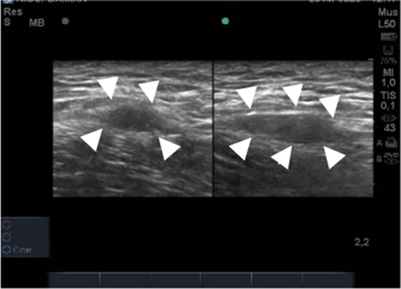
The US examination of the main body of the proximal tendon or “non insertional” part (above to greater trochanter) demonstrated an increased size of the tendon. In one of the cases (39 yrs.) a large increased size was demonstrated, and the other patient (30 yrs.) showed a focal spindle or fusiform shape as a focal tendinopathy with hipoechogenicity appearance (Figure 1).
Discussion The sonographic appearance of non-insertional TFL tendinopathy we describe is the same that has been described for the main lower limb tendinopathies (Achilles, patellar or plantar fascia).
In our opinion both, Sonographic and MRI are very operator dependent and in this two cases required a meticulous scanning technique to visualize the non-insertional tendinopathy. Point tenderness in proximal TFL has been described in US as a possible insertional tendinopathy, but the “non insertional” proximal TFL tenderness has not been described up until yet in US examination.
US examination of TFL must include the “non-insertional” area of the tendon proximal to greater trochanter of femur. These two cases illustrate an undescribed and atypical presentation of TFL tendinopathy.
Transverse and longitudinal image of TFL
References 1 Bancroft LW, Blankenbaker DG. Imaging of the tendons about the pelvis. AJR 2010; 195: 605–617.
2 Bass CJ, Connell DA. Sonographic findings of tensor fascia lata tendinopathy: another cause of anterior groin pain. Skeletal Radiol 2002.31:143–148.
3 Bossy M, Pedret C. Entesopatía del tensor de la fascia lata. Apunts Med Esport. 2013; 48(178):77–80.
4 Jiménez Díaz JF, Alvarez Rey G, Balius Matas R. New technologies applied to ultrasound diagnosis of sports injuries. Adv Ther. 2008; 25(12):1315–30.