Article Text

Abstract
Background: In football there are established age-related tournaments for males and females to guarantee equal chances within the game for all the different age groups. To prevent participation in the incorrect age group, and owing to the fact that in some Asian and African countries registration at birth is not compulsory, other methods of age determination need to be available. Standard radiographs of the left wrist have been used for assessment of skeletal age for many years. This is, however, not ethical in the sporting environment.
Aim: To study the possible use of magnetic resonance imaging (MRI), which has no radiation risk, in estimating the age of healthy adolescent football players.
Methods: The examination protocol was applied in four countries using, their respective MRI equipment using a 1-T or 1.5-T magnet and a wrist coil. 496 healthy male adolescent football players between the ages of 14 and 19 years from Switzerland, Malaysia, Algeria and Argentina were selected for the study. The degree of fusion of the left distal radial physis was determined by three independent raters by a newly developed grading system which can be used in future MRI epiphysial fusion grading studies.
Results: The inter-rater reliability for grading was high (r = 0.91 and 0.92); all correlations were highly significant (p<0.001). The average age increased with a higher grading of fusion, and the correlation between age and grade of fusion was highly significant (r = 0.69, p<0.001). Only one player (0.8%) in the 16-year-old age group was graded as completely fused.
Conclusion: MRI of the wrist offers an alternative as a non-invasive method of age determination in 14–19-year-old male adolescents. The grading system presented here clearly identifies the skeletal maturity by complete fusion in all MRI slices, which eliminates any risk associated with standard radiographic rating as determined by the International Atomic Energy Agency.
- MRI, magnetic resonance imaging
- PACS, picture archiving and communications system
Statistics from Altmetric.com
In football there are established age-related tournaments for males and females, to guarantee equal chances within the game for all the different age groups. Over the years, the tournaments have gained momentum and popularity, particularly the under-17 competitions. Unfortunately, there has been some suspicion that the biological age of the participating players might be older than the documented age as stated by the passport or birth certificates used to determine the eligibility of the individual. This situation is aggravated by the fact that in some Asian and African countries registration at birth is not compulsory. Thus, reliable methods for proper age estimation are required. Discrepancies in age lead to unequal chances and are against the spirit of the game and, of course, “fair play”. Malina et al1 examined maturity-associated variation in sport-specific skills of youth soccer players, concluding that age, experience, body size and stage of puberty contribute considerably in different combinations to the variance of some football skills such as dribbling with a pass, ball control with the body and shooting accuracy. Also, players with a greater relative (or possibly false lower) age are more likely to be identified as “talented” because of the likely physical advantages they have over their “younger” peers.2
Standard radiographs of the left wrist for assessment of skeletal age have been described by Todd,3 Greulich and Pyle,4 Tanner,5,6 and earlier using the Fels method,7 which is still widely used for assessment of skeletal age. The determination of skeletal maturity has an important place in the practice of paediatrics, especially in relation to endocrinological problems and growth disorders. Furthermore, it is essential in any attempts (perhaps misguided) at treating short children with anabolic steroids.8
Standard radiographs are also used medicolegally to determine age in a court of law. Their use is based on a court order by a judge who allows the use of the limited radiation to obtain the images needed to determine the skeletal age. The International Atomic Energy Agency regulates the use and possible abuse of x rays under the title “International basic safety standards for protection against ionising radiation and for safety of radiation sources” (CD-ROM edition, 2003, Geneva, Switzerland). Thus, in sports the use of x rays (a radiation exposure) to determine players of over age is not allowed by a court of law as the action does not amount to criminal action. In addition, skeletal age determined on standard radiographs may vary depending on ethnic origin (table 1).
Summary of assessed studies on ethnic differences in skeletal age and chronological age
As the screening of football populations by using radiographic examination cannot be justified, other methods such as ultrasound have been investigated9; however, only children up to 6 years were examined. Age-related values are not available.
Such an x ray-free examination method could be used in all sports in case of discrepancies or suspicion that a date of birth presented is inappropriate. This might be of interest not only to football but also to the entire sports community and the International Olympic Committee, where age determines competing categories, as well as for paediatricians, when dealing with endocrinological or other disorders, and also for courts, when dealing with criminal offence by under-aged persons.
Aims of the study
-
To develop a grading system of magnetic resonance imaging (MRI) for epiphysial fusion of the distal radius
-
To evaluate the reliability and validity of the grading system of MRI examinations of the wrist to determine skeletal bone age in the age group 14–19 years
-
To compare scores of the male football players from different ethnic groups
METHODS AND POPULATION
Examination protocol
The examination protocol was applied in all centres (Zurich, Kuala Lumpur, Buenos Aires, Algiers) using their respective MRI equipment with a 1-T or 1.5-T magnet and a wrist coil.
The wrist was positioned above the head or at the side of the body. The third metacarpal was placed as close as possible to the same axis as the radius.
Coronal sequences were planned parallel to the distal velar radial surface.
The imaging parameters were not pushed to the level of the top of the line magnets in order to allow protocol transfer to the equipment available worldwide. The numbers are based on Siemens equipment. The values may be slightly adapted for other manufacturers.
The following parameters were applied: T1-weighted spin echo, TR 350–500 ms, TE 12–20 ms, slice thickness 3 mm, interslice gap 0.3 mm (1.1 distance factor), pixel size ⩽0.5 mm (eg 12 cm field of view with a 256 matrix), 2–4 acquisitions and 9 images (to cover the entire distal radius from anterior to posterior).
Nine images were printed per film, with the wrist enlarged so as to include the distal 3 cm of the radius, and the entire carpus was included. Where available the images were saved to allow evaluation on a picture archiving and communications system (PACS).
Grading system
In a pilot study, the authors reviewed conventional radiographs of the left wrist from a normal hospital population and compared them with MRI of the left wrist of age-matched healthy volunteers. Grading parameters were agreed upon after statistical analysis of inter-rater reliability and correlation with the chronological age. Box 1 and figs 1–6 present the classification for grades I–VI for fusion of the physis of the distal left radius.

Grade I; T1-weighted spin-echo images of completely unfused distal left radius (magnification on the left, original image on the right).
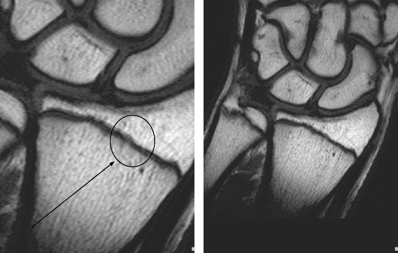
Grade II; T1-weighted spin-echo images of early fusion of distal left radius showing minimal hyperintensity within the physis (circle). (Magnification on the left, original image on the right.)

Grade III; T1-weighted spin-echo image of distal left radius showing trabecular fusion of <50% of the radial cross-sectional area. (Magnification on the left, original image on the right.)

Grade IV; T1-weighted spin-echo image of distal left radius showing trabecular fusion of >50% of the radial cross-sectional area. (Magnification on the left, original image on the right.)

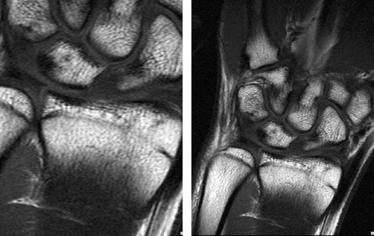
Grade V; T1-weighted spin-echo image of distal left radius showing residual physis <5 mm on any one section. (Magnification on the left, original image on the right.)

Grade VI; T1-weighted spin-echo image of complete fusion of distal left radius. (Magnification on the left, original image on the right.)
Box 1: Classification criteria for ossification/fusion of the distal radius on magnetic resonance images
-
Grade I: Completely unfused
-
Grade II: Early fusion: minimal hyperintensity within the physis
-
Grade III: Trabecular fusion of <50% of the radial cross-sectional area
-
Grade IV: Trabecular fusion of >50% of the radial cross-sectional area
-
Grade V: Residual physis, <5 mm on any one section
-
Grade VI: Completely fused
The raters (three of the four authors) were blinded to the name, age and country of origin. Two of them were experienced radiologists and one a specialist in neurology. The blinding code was prepared by the fourth author (epidemiologist). The three individual gradings were computed to a majority grading using the most common grading or if all three ratings deviated, the average grading.
Statistical analysis
All data were processed on a Macintosh computer (Apple Computer, Cupertino, California, USA) using Microsoft Excel (Microsoft Corporation, Redmond, Washington, USA). The statistical procedures were performed using StatView V.5.0. Statistical methods applied were frequencies, cross tabulations, descriptives and means. Depending on the type of data, correlations were analysed using Pearson’s coefficient of interval data and Spearman’s (r) rank correlation for ordinal data. Differences between the groups were examined using either the Student’s t test or the Wilcoxon’s signed rank test for dependent pairs or the Kruskal–Wallis H test. Significance was accepted at the 5% level.
Population
To account for ethnic differences, healthy male adolescents from Switzerland, Malaysia, Algeria and Argentina were selected under the condition that there is absolute certainty concerning birth certificate issued by governmental institutions. Young healthy male football players aged between 14 and 19 years were selected by the respective national football association or by regional football clubs. Exclusion criteria were previous fracture of the forearm or wrist and endocrinological or other systemic disorders. Ethical approval for the study was obtained by the respective national institution, and informed consent was obtained according to local ethics committee recommendations.
In total, 496 boys were examined in four different countries (table 2). The players were grouped according to their age. Age group was calculated (date of MRI minus date of birth, eg the group of 14-year-olds was defined as having had their 14th birthday but not their 15th birthday, the group of 16-year-olds are all between 16 and 17 years for 17th birthday minus 1 day).
Number of participants in different age groups and countries
RESULTS
Reliability of the rating
The reliability of the ratings was evaluated by analysing inter-rater reliability, intra-rater reliability (test–retest) and a comparison of ratings obtained from PACS and hard copies of MRIs.
Inter-rater reliability
Three raters independently graded MRI hard copies of the 471 cases (in 25 cases one rating was missing but the other two ratings were identical). In 218 cases all three raters agreed on an identical grading (46%), and in further 244 (52%) cases two of the raters agreed. Thus, in only 2% was no agreement observed between the raters. In 97.7% the gradings were either identical or deviated by one category; in only 11 cases (2.3%) was the range between the three ratings two categories. The inter-rater reliability (r) ranged between 0.91 and 0.92. All correlations were highly significant (p<0.001). The agreement of the three individual raters with the majority grading was even higher (r = 0.95–0.97).
Test–retest reliability
For analysis of test–retest reliability, a mixed sample of 96 Swiss and Malaysian cases (hard copy and PACS) was analysed twice within the same day. The individual raters had an identical grading in 83–86% of the cases. The intra-rater reliability (r) ranged between 0.96 and 0.98. All correlations were highly significant (p<0.001). Most grading was identical in almost all cases (n = 90; 94%); in only six cases did it deviate by one category. Thus, no significant difference was observed in the average majority grading,
Comparison of PACS and hardcopies
Using the subgroup of 111 Swiss cases, the gradings based on PACS and hard copies were compared. The gradings of the individual raters all correlated highly significantly (r = 0.9–0.94, p<0.001) and so did the majority rating (r = 0.94, p<0.001) between the two methods. No significant difference was observed in the average majority gradings (Wilcoxon’s test, p = 0.88). In most cases (n = 80; 72%), the majority gradings were equal for both methods. In 28% (n = 31) of cases they deviated by one category.
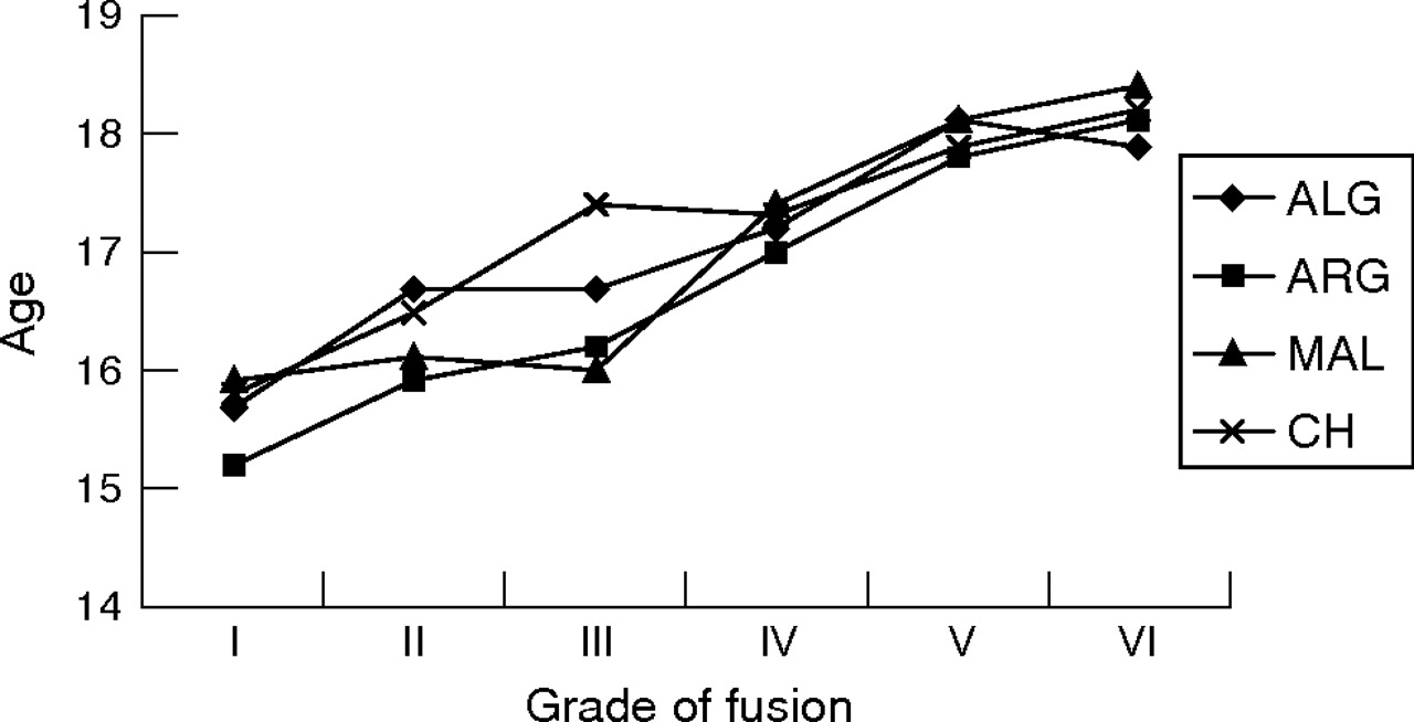
Relationship between age and grading of fusion
Table 3 presents the grading of fusion in relation to age. The average age increased with a higher grading of fusion (table 4 and fig 7). The correlation between age and grade of fusion was highly significant (r = 0.69. p<0.001). Only one player (0.8%) in the 16-year-old group was graded as completely fused (from the Malaysian group).
Number (%) of participants in relation to grading of fusion in the different age groups
Number of participants and average age in different categories of fusion grading

Box plot of grade of fusion by age at magnetic resonance imaging (MRI).
Table 5 and fig 8 show the comparison of the MRI gradings among the four countries examined.
Mean (SD) age-dependent grading of fusion in different countries
Comparison of the magnetic resonance imaging rating for the four examined countries. ALG, Algeria; ARG, Argentina; CH, Switzerland; MAL, Malaysia.
DISCUSSION
The determination of skeletal maturity has an important place in the practice of paediatrics, especially in relation to endocrinological problems and growth disorders. Age is also decisive for the punishment of delinquents in a court of law. In sport, in particular football, competitions have been designed according to age groups to guarantee equal chances within the spirit of “fair play”. Standard radiograph analyses of the left wrist have been used for decades to estimate the age and potential to grow following the published standards established by Greulich and Pyle,4 Tanner5 and Fels.7 The change in socioeconomic factors, in the environment and possibly in nutritional habits has influenced the comparison of standards with current radiographic assessment. Ethnic differences unrelated to these changes also have also been shown by several authors and also with controversial results for the same ethnic group.
Todd, Greulich and Pyle together designed a long-term investigation of human growth and development in 1929. The study commenced in 1931, examining children at 3-month intervals for the first postnatal year, at 6-month intervals from 12 months to 5 years and annually thereafter until age 18 years. Radiographic films were made of the left shoulder, elbow, hand, hip, knee and foot. A thousand children were included in the study in the Cleveland area of the USA, and the results served as a source of information for the Radiographic atlas of skeletal development of the hand and wrist.4 The authors presented age, and gender-related standards to be used for comparison. The standard deviation for the skeletal age of 17-year-old boys was 13 months and that for 16-year-old girls was 7.31 months. Skeletal maturity—that is, complete fusion of the wrist bones—has been observed at age 18 years in boys and at 17 years in girls by Tanner5; later, Tanner and Whitehouse presented standards from birth to maturity by using x ray and including other parameters such as height, weight, and height and weight velocity to obtain a mathematical formula to calculate maturity.6 The methods of examination and assessment have been re-evaluated and compared and show good correlations using regression analysis.10 However, on applying scatter plots instead of regression analysis, the difference between the two methods shows an unacceptable error for clinical practice. The authors recommended the more time-consuming TW211 method for assessing skeletal age.
The original methods involved North American and UK children and young adolescents to establish the normative values; however, the question of ethnic differences has been raised by several authors. The European population in Denmark,12 Spain13 and Holland14 presented good correlation with Greulich and Pyle and Tanner standards; Turkish boys,15 however, advanced in their skeletal age faster. The South American16 population presented good correlation using the TW2 technique, whereas a sample in sub-Saharan Africa17 showed slower skeletal age development. In China and Japan, faster maturity has been observed in comparison with the European population.18–20 Studies from the USA present controversial observations.21–26 Loder observed faster maturation in black and white boys and girls when compared with G&P standards; Ontell described faster maturation in black and Hispanic girls and black and Asian boys, with white boys trailing in skeletal maturity. On the contrary, Mora found faster skeletal maturation in European Americans when compared with African Americans (table 1).
The need for an alternative method of determining age and maturity has been raised by the International Atomic Energy Agency regulatory body, which does not allow x ray examination except when clinically justified for the individual, which is not the case for age determination in sports or even in medicolegal situations except when a court order exists, based on criminal charges.
What is already known on this topic
-
Standard radiograph of the left hand and wrist is currently used in skeletal age assessment methods.
-
The appearance of distal radial growth plate fusion using standard radiographs.
What this study adds
-
The appearance of different degrees of fusion of the distal radius epiphysial growth plate using magnetic resonance imaging (MRI), a radiation-free imaging modality that can be used in healthy people such as athletes and football players.
-
An MRI grading system for the different degrees of fusion of the distal radius growth plate, which can also be used for other growth plate fusion studies with high intrarater and inter-rater reliabilities.
-
The significant correlation between age and MRI grade of fusion of the distal radial growth plate.
MRI offers an alternative as a non-invasive method of examination. The grading system presented clearly identifies different degrees of epiphysial fusion of the distal radius. Inter-rater and intrarater reliabilities are high and the learning curve steep because of clear and simple criteria, even for a non-radiologist. Complete fusion occurs at the age of 17–18 years in the ethnic groups examined, with faster maturation among Argentinian and Malaysian boys in comparison with Algerian and Swiss. The mean age of participants with complete fusion of the radius was 18.3 years (SD 0.9) indicating that complete fusion is very unlikely to occur at 17 years of age. In our population only one boy out of 130 aged 16 (0.8%) presented complete fusion. Most boys in the age group between 16 and 17 years presented as grade II (table 3). The current data justify extension of the examined population to other ethnic groups such as sub-Saharans, East Asians and Central Americans. As the presented study did not register weight and height, we recommend including anthropometrical data including body mass index to analyse the possible influence on the speed of maturation.
In conclusion, MRI offers an alternative as a non-invasive method of examination of epiphysial fusion. The grading system can accurately identify the variable degrees of epiphysial fusion in an objective teachable manner.
REFERENCES
Footnotes
-
Published Online First 4 October 2006
-
Competing interests: None.
-
Ethical approval: Ethical approval for the study was obtained by the respective national institution, and informed consent was obtained according to local ethics committee recommendations.