Article Text

Abstract
Objectives To study fetal wellbeing and uteroplacental blood flow during strenuous treadmill running in the second trimester.
Methods Six pregnant Olympic-level athletes in endurance events aged 28–37 years and training 15–22 h per week before the pregnancy were tested once at 23–29 weeks of pregnancy. The women ran three to five submaximal workloads on a treadmill with approximately 60–90% of maximal oxygen consumption. The maternal–fetal circulation was assessed with Doppler ultrasound of the uterine and umbilical arteries before, during and after exercise.
Results Mean uterine artery volume blood flow was reduced to 60–80% after warming up and stayed at 40–75% of the initial value during exercise. Fetal heart rate (FHR) was within the normal range (110–160 bpm) as long as the woman exercised below 90% of maximal maternal heart rate (MHR). Fetal bradycardia and high umbilical artery pulsatility index (PI) occurred when the woman exercised more than 90% of maximal MHR and the mean uterine artery volume blood flow was less than 50% of the initial value. FHR and umbilical artery PI normalised quickly after stopping the exercise.
Conclusions Exercise at intensity above 90% of maximal MHR in pregnant elite athletes may compromise fetal wellbeing.
Statistics from Altmetric.com
Exercise of moderate intensity is safe during uncomplicated pregnancy.1,–,3 The American College of Obstetricians and Gynecologists4 states that a woman with a low-risk pregnancy can participate in moderate physical activity for more than 30 min per day. More vigorous exercise is also regarded as safe for women who are well trained before the pregnancy.5,–,8 A more controversial issue has been intensive training during pregnancy. Some elite athletes plan to deliver a baby in between competitions such as the Olympics or world championships. They want to maintain a high level of performance throughout pregnancy.
In uncomplicated pregnancy adverse effects of training on the fetus are unlikely.9 Most studies show a small or moderate increase of the baseline fetal heart rate (FHR) during or after maternal exercise. FHR decelerations and bradycardia are uncommon.7 9 10 Studies of possible effects of strenuous exercise on uteroplacental blood flow are sparse. The number of pregnant women who engage in training during pregnancy is growing, and there is a need for establishing intensity values above which exercise may be harmful for the fetus.
A common and simple way to monitor training intensity is to record maternal heart rate (MHR) during exercise. Target MHR zones and guidelines for exercise during pregnancy have been published.11 12
Volume blood flow to the pregnant uterus during exercise has been sparsely studied. One experimental study in pregnant sheep found reduced volume blood flow by 15–20% in response to different exercise regimens.13 Studies in pregnant women have produced contradictory results.14,–,16 There are several Doppler ultrasound studies on exercise-related changes in umbilical and uterine artery waveforms,17,–,20 but we have found no studies on uterine artery volume blood flow during strenuous exercise in pregnancy.
The primary aim of the present study was to examine the effects of strenuous treadmill running on fetal wellbeing in pregnant elite athletes. The secondary aim was to assess volume blood flow to the pregnant uterus during intensive exercise.
Methods
Seven pregnant athletes representing Norwegian national teams in endurance events (cross-country skiing, duathlon, long distance running and race walking) were invited to participate in the study from October 2002 to March 2006. Women were between 28 and 37 years of age and all but one were nulliparous. In the non-pregnant state they trained from 15 to 22 h per week, and all together they had won 21 medals in Olympic and world championships. They had been tested using the same test personnel on several previous occasions at the Olympic training centre or the Norwegian School of Sports Science. Written informed consent was obtained before inclusion. The study protocol was approved by the Regional Committee for Medical and Health Science Research Ethics in Southern Norway.
One athlete was tested twice (at 15 and 24 weeks of pregnancy), but she was not included in the study. She was the first athlete of the test series, and her results were used to pilot the study protocol. The study population thus consisted of six women tested once at 23–29 weeks of pregnancy. All women had a dating scan at around 18 weeks of pregnancy. Before the treadmill test the women were examined with ultrasound to assess normality regarding fetal growth, amniotic fluid volume, blood flow in the uterine arteries and cervical length. None of them had ‘notch’ or high mean pulsatility index (PI) of the uterine arteries before the treadmill test.
Exercise testing
After warming up for 10 min with MHR of approximately 135 bpm (Polar Vantage, Polar Sports Tester, Kempele, Finland), the women ran three to five submaximal workloads on a treadmill (Woodway, Wurzburg, Germany) with a range of approximately 60–90% of maximal oxygen consumption (Vo2). The speed was increased in steps of 1 km/h, and the inclination of the treadmill was 6% throughout the entire test. Each workload lasted 5 min (in order to reach a Vo2 plateau), and Vo2 was measured during the last minutes using a nose-clip and a mouthpiece (Hans Rudolph, Hans Rudolph Inc, Kansas City, USA) with a one-way valve connected to an automated gas exchange analyser (Oxygen Champion; Jaeger, Wurzburg, Germany). Between each bout there was a 4-min pause for fingertip sampling of capillary blood (50 ml) used for the determination of blood lactate concentration (1500 Sport Lactate analyzer; YSI, Yellow Spring, USA) and ultrasound assessments of the maternal–fetal circulation. Each athletes' prepregnant maximum heart rate was adjusted for the known increased maximum heart rate during pregnancy.12 Blood lactate was analysed after the treadmill test and not used as a stopping rule for further testing.
Ultrasound
We assessed the uteroplacental blood flow with Doppler ultrasound before, during and after the exercise tests with a Logic 7 device (GE Vingmed Ultrasound, GE Healthcare, Norway) with a multifrequent curvilinear transducer 3–7 MHz. The women were in the semirecumbent supine position during the ultrasound examinations. We assessed PI and FHR in a free loop of the umbilical artery. Umbilical artery PI should not exceed 1.5 (mean±2 SD for 23–29 weeks) throughout the test.21 The normal FHR range was defined as 110–160 bpm. Fetal tachycardia (FHR >160 bpm) during the treadmill test was accepted, but fetal bradycardia (FHR <110 bpm) immediately after one workload was regarded as a stopping rule for further testing.
Uterine arteries were assessed on both sides at the level of the crossing of the internal iliac arteries. We used colour Doppler to identify the uterine artery and to optimise the insonation angle (kept at <30°). The uterine artery diameter was measured in B mode three times on each side before the treadmill test. Calipers were placed on the inner vessel wall, and the mean diameter was used throughout the treadmill test. The volume blood flow of both arteries was calculated automatically by the machine software during pulsed Doppler measurements. TAMEAN was calculated as: TAMEAN=sum{Vmean} from t1 to t2/(t2−t1) (cm/s). VolFlow (ml/min)=area (cm2)*TAMEAN (cm/s)*60 (s) where the area was calculated from the vessel diameter (3.14×diameter/2)2.
The pause between workloads was 4 min (1 min to shift from the treadmill to supine position and back again and 3 min for measurements). After each workload as many measurements as possible were recorded. The right and left uterine arteries and the umbilical artery were measured in a repeated sequence with at least two complete recordings from each vessel within the 3-min interval. The first FHR and umbilical artery PI value was used. For the uterine artery the technically best recording on each side within the interval was used. The mean volume flow in the resting state before the start of the treadmill test was set to 100%, and the proportional change from the initial value was calculated. The final measurements were performed after 10 min rest.
Results
FHR was within the normal range as long as the mother exercised below 90% of maximal MHR (figure 1). Fetal bradycardia occurred when the mean uterine artery blood flow was less than 50% of the initial value (table 1) and the exercise intensity was greater than 90% of maximal MHR (figure 1).

Plot of fetal heart rate (FHR) versus maternal maximal capacity (% of maximal maternal heart rate; MHR). Shaded area is the normal FHR range (110–160 bpm).
Maternal and fetal parameters before, during and after exercise
Four women completed three workloads uneventfully and stopped themselves from further exercise. One woman (case 3) had estimated volume flow in the uterine arteries of 41% of the initial value after the final workload, and maternal blood lactate was 4.5 mmol/l. She had exercised at 88% of maximal MHR, and the fetus showed no sign of compromise. One woman (case 5) completed four workloads. Immediately after the fourth workload, the fetus had bradycardia (103 bpm) and high umbilical artery PI (1.67). Further exercise was stopped. At this point the estimated volume flow in the uterine arteries was 37% of the initial value, and she exercised at 97% of maximal MHR. Maternal blood lactate was 4.9 mmol/l. The fetus recovered quickly with no signs of sustained bradycardia or high PI in the following 10 min of rest. One woman (case 6) completed five workloads. This fetus also had bradycardia (92 bpm) and high umbilical artery PI (1.65) immediately after the last workload. The volume flow was estimated to be 42% of the initial value, and the maximal MHR was 92%. Maternal blood lactate was 3.0 mmol/l. Both FHR and umbilical artery PI normalised quickly when exercise was stopped.
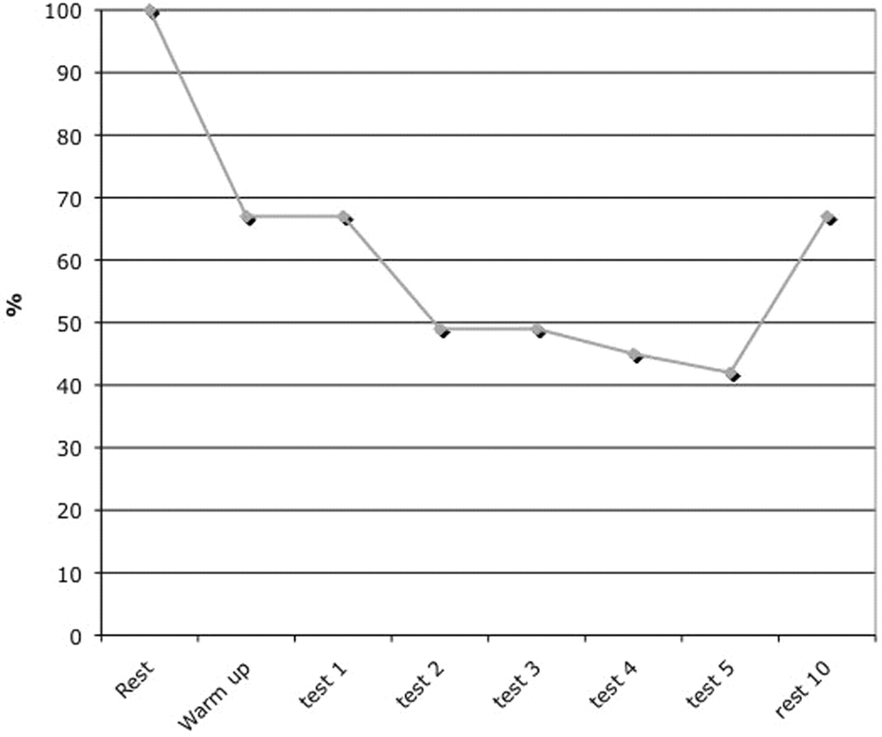
The mean uterine artery volume flow was reduced to 60–80% of the initial value in all women after warming up and before the test series itself started (table 1). Throughout the test series the volume flow stayed at approximately 40–75% of the initial values. After 10 min of rest the mean volume blood flow was back to approximately 90% of the initial value in four women. In two women the recovery of blood flow was somewhat slower (table 1). Figure 2 shows the volume blood flow profile (percentage of initial value) during exercise for case 6. The other women had similar flow profiles.
Proportional change of mean uterine artery blood flow during the treadmill test for case 6.
Some maternal characteristics and pregnancy outcomes are given in table 2. Case 6 developed haemolysis–elevated liver enzymes–low platelets syndrome at 35 weeks. She did not have pre-eclampsia in her first pregnancy. Labour was induced, and she delivered a healthy girl vaginally with a birth weight of 2285 g. The other women delivered between 36 and 42 weeks, and the babies weighed 3000–3440 g (table 2). Birth weight related to gestational age was in the lower normal range (86–103%) for Norwegian children (table 2). There were four normal deliveries, one caesarean section and one vacuum delivery. The two operative deliveries were caused by obstructed labours.
Maternal characteristics and pregnancy outcomes
Discussion
The present study suggests that fetal wellbeing may be compromised during strenuous exercise among pregnant elite athletes. Exercise intensity should not exceed 90% of maximal MHR when there is simultaneous reduction in uterine volume blood flow to less than 50% of the initial value. However, the sample size was small, and the results should be viewed with caution.
Blood lactate after the last workload was 3.0 mmol/l or greater in four out of six women (table 1), but only the two women with simultaneous maximal MHR greater than 90% and uterine volume blood flow less than 50% experienced fetal bradycardia and high umbilical artery PI. We postulate that maximal MHR and uterine volume blood flow may be more important parameters than blood lactate in defining a safety zone for fetal wellbeing during strenuous exercise.
Uterine artery volume blood flow in pregnant elite athletes during exercise has been rarely studied, but the non-invasive measurement of uterine artery volume flow has been validated.22,–,24 Palmer et al22 found that it is feasible to estimate uterine artery volume blood flow in pregnant women using pulsed-wave Doppler. Acharaya et al23 validated experimentally (in sheep) that the blood flow can be measured with reasonable accuracy, and that the non-invasively measured flow correlates well with the directly measured flow. Rigano et al24 also showed that it is feasible to measure uterine artery volume blood flow using ultrasound. However, unlike the present study, the last two papers measured the diameter of the vessel using power Doppler angiography rather than B-mode ultrasound.
In the present study, volume blood flow was calculated automatically by the machine software with the use of TAMEAN values and the area of the vessel lumen (3.14×(diameter/2)2). Any measurement error of the vessel diameter will have a huge impact on the flow in ml/min. Vessel diameter was assessed as the mean of three B-mode measurements. The use of power Doppler angiography mode may have improved the measurements.25 For time reasons we did not measure the vessel diameter after each workload, but postulated that the vessel diameter remained unchanged during exercise. This is in accordance with the results from a study by Jeffries et al.16 If the uterine artery diameter did change during exercise, the flow would be different from what we have estimated. However, for a repeated measurement design a systematic error in the measurement would not matter a great deal because we were interested in the changes over time rather than in the absolute flow values. The proportional change in volume flow in the same individual should be a more robust parameter than volume flow in ml/min.
Changes in the mean volume blood flow during the treadmill tests were almost identical to the results from a study of pregnant sheep by Lotgering et al.13 They found a rapid reduction in volume blood flow to 80–85% of the initial value when pregnant sheep exercised on a treadmill.13 The reduction in volume flow during exercise was higher in athletes in the present study than among sheep. Whether this difference is species related or exercise-intensity related is not known.
Previous studies in pregnant women have produced contradictory results. Rauramo and Forss14 found unchanged uterine blood flow following exercise. However, the study used Xenon clearance techniques, and that methodology was probably not reflecting uterine artery volume flow. One study of portal vein blood flow found that treadmill exercise decreased splanchnic blood flow by more than 50%.15 The authors argued that reduced splanchnic blood flow reflects reduced uterine blood flow. If this is correct, the results are in line with our study. Jeffreys et al16 examined blood flow in the right uterine artery in supine and left lateral positions during rest and exercise in the second trimester. It is difficult to compare our results with that study because only one uterine artery was examined. Both arteries should be examined when total or mean volume blood flow to the uterus is estimated.
Measurement of uterine artery volume blood flow is time consuming and difficult to do. MHR recording during exercise is a simple way to monitor training intensity and can be done with a HR monitor. Target MHR zones and guidelines for exercise during pregnancy have been published.11 12 We found that exercise at less than 90% of maximal MHR may be regarded as a safety zone for elite athletes. However, the American College of Obstetricians and Gynecologists no longer recommend MHR targets to assess the intensity of exercise, but prefer self-regulation and scales of perceived exertion.26 Elite athletes have trained for many years and will usually have tested maximal Vo2 on several previous occasions in the non-pregnant state. They should be able to identify thresholds and self-regulate training intensity without Doppler ultrasound and MHR recordings.
What is already known on this topic
Exercise of moderate intensity is safe during uncomplicated pregnancy.
What this study adds
▶ Exercise at intensity above 90% of maximum maternal heart rate may induce fetal bradycardia.
▶ Mean uterine artery volume blood flow was reduced by 25–60% during intensive exercise.
A limitation of the present study is the small sample size. The number of eligible women was limited because we only included pregnant Olympic-level athletes in endurance events. We do not know if the study results can be generalised to other groups of women who may be physically fit and train regularly, but do not have ‘elite’ status. Several studies have demonstrated that less vigorous exercise in low-risk pregnancies is safe for the mother and fetus.1,–,8 More studies of both elite athletes and physically active pregnant women are needed to establish intensity values above which exercise is potentially harmful to the fetus.
The present study was not designed to assess long-term outcomes after intensive training during pregnancy. One study has indicated reduced birth weight and thinner babies in women who were randomly assigned to a high volume of exercise in mid and late pregnancy.27 Birth weights for gestational age of the children in our study were in the lower normal range for Norwegian children (86–103%). We were unable to draw any conclusions based on the small sample size.
An interesting research question would be to assess volume blood flow in the uterine arteries and MHR/FHR recordings during exercise tests in pregnancies with uteroplacental insufficiency. Exercise tests in pregnant women with uteroplacental insufficiency could be done with initial low intensity workloads, and then increase the intensity stepwise under careful control of the mother and the fetus. Results from previous studies of uterine artery and umbilical artery waveforms have been contradictory. One study found a transient deleterious effect in a subset of women with uteroplacental insufficiency after submaximal exercise,19 whereas another study concluded that maternal exercise did not significantly alter uterine and umbilical perfusion in normal or growth-restricted fetuses.20
We observed that fetal bradycardia and high umbilical artery PI could occur when women exercised at intensities greater than 90% of maximal MHR with simultaneous reduction in uterine volume blood flow to less than 50% of the initial value. Therefore, we conclude that exercise at intensity above 90% of maximal MHR in pregnant elite athletes may compromise fetal wellbeing.
References
Footnotes
-
Competing interests None.
-
Patient consent Obtained.
-
Ethics approval This study was conducted with the approval of the Regional Committee for Medical and Health Science Research Ethics in Southern Norway.
GE Healthcare Norway provided the ultrasound device (Logic 7) free of charge. GE had no influence on writing the study protocol, analysing the data or interpreting the results.
-
Provenance and peer review Not commissioned; externally peer reviewed.