Article Text

Statistics from Altmetric.com
The role of the team physician for intercollegiate ice hockey teams is unique. Owing to the rather diverse characteristics of many of the mechanisms of injuries for on-ice activities, it is important that team physicians have a good knowledge of the fundamentals of the game in order to understand when a player can return to on-ice activities. It is also essential that the team physician understands the underlying passion in the great majority of players for returning to competition. Unlike many other sportsmen, ice hockey players have a strong desire to return to competition, even after fractures or dislocations that would make athletes in other sports be sidelined for long periods of time. Therefore, in addition to understanding the on-ice activities, it is important that the team physician understands the individual athletes, either personally or through his/her relationship with the team’s athletic trainer, to determine a safe time for these athletes to return to competition. The purpose of this review is to summarise the main injuries seen in high level intercollegiate ice hockey and to review the practical treatment problems for physicians who provide coverage for high level amateur hockey teams.
Aetiology of ice hockey injuries
The vast majority of division I ice hockey injuries occur during games. In fact, the injury rate in practices of 2.1/1000 athlete exposures is one of the lowest in intercollegiate sports.1 However, the game injury rate of 17.5/1000 athlete exposures is one of the highest among team sports.1 Therefore on-site coverage of most division I and high level ice hockey games is necessary.
There is a distinct distribution of the types of injury that occur in ice hockey. Firstly, early season injuries are almost always overuse or sprain and strain type injuries. Groin, or adductor, strain is one of the most common lower body injuries, but almost always occurs within the first few months of the season. Concussions are becoming one of the more common time-loss injuries.1 These can occur throughout the season, but they rarely occur in practice. The other two common time-loss injuries are medial ligament injuries of the knee and acromioclavicular (AC) joint separations of the shoulder.
Most injuries that occur in intercollegiate ice hockey do not require surgery. In fact, the rate of surgery per player is the second lowest of the 28 sports tracked by the National Collegiate Athletic Association (NCAA) database.1 Therefore a broad-based knowledge of the most common ice hockey injuries is necessary for a team physician. Early recognition of injuries can help to minimise downtime, with earlier return of athletes to competition. Therefore it is important to understand prevention and early diagnosis and treatment of these injuries and specific tricks that can be used to return an athlete sooner to on-ice activities. An overview of the most common ice hockey-specific injuries is covered in the following sections.
Early season groin strains
One of the most common early season injuries is hip adductor, or groin, strain.1 This is also one of the most common injuries in the NCAA and National Hockey League ice hockey injury databases. Groin strains almost always occur as the result of a sudden change in training habits. In the majority of cases, this occurs at the start of the season, with the initiation of a hard, on-ice training regimen. Pain from a groin strain is almost always localised to the pubic part of the pelvic bone or at the adductor musculotendinous junction. The pain associated with adductor strains is almost always increased with any manoeuvre in which the thigh crosses the midline of the body, especially with power skating and cross-over skating.
In most cases, groin strains are a self-limiting condition, which resolve with time. However, because they almost always occur during the early part of the season, including during try-outs, the problem can seem to last an eternity to some players and coaches and can affect whether the player makes the team or not and which lines or specialty teams they play on. For minor injuries, a good stretching and warm-up programme, followed by avoidance of on-ice activities that cause symptoms, usually results in pain resolution over the course of a few days. Players should avoid significant power skating or cross-overs until the symptoms have completely resolved. Over-the-counter anti-inflammatory drugs can also be useful for decreasing some of the pain caused by inflammation. It is important that these drugs be used with an appropriate stretching and warm-up programme.
For groin strains that prevent skating because of pain, a programme of rest, ice and ultrasound is recommended until the athlete can resume skating. It is important that athletes cross-train using cycling or pool therapy to try to maintain their cardiovascular status at a high level until the symptoms improve. In cases in which symptoms do not resolve over a week or two, further investigation should be performed to determine if the pain is from another source, such as sports hernia or femoroacetabular impingement.
Overall, one of the best ways to treat groin strains is to work on prevention. Players should work with their strength coach and athletic trainer on a programme of stretching and hip adductor strengthening before the initiation of any on-ice activities, so that they do not develop overuse injuries of the groin. It is especially important to perform a stretching programme after appropriately warming-up. A short (500 m) jog or a few laps around the ice may be necessary to stimulate the blood circulation to the muscles so that the player can stretch appropriately.
It goes without saying that, if the symptoms from a groin strain linger for more than 2–3 weeks, an athlete should be further evaluated for sports hernia or femoroacetabular impingement. An examination to determine if they have pain when performing an abdominal crunch or a partial sit-up (which would point towards a sports hernia) or have pain on hip flexion and internal rotation (which would point to a posterosuperior acetabular labral tear) should also be part of the initial evaluation of athletes with groin strains.
Skate (lace) bite
“Skate bite” or “lace bite” is a relatively common ice hockey injury. It almost always occurs early in the hockey season in players with new skates or inflexible older skates. Skate bite is almost always caused by too much pressure from a new stiff skate tongue, which has not been broken in well, or in old skates that have old and inflexible skate tongues. In both situations, the inflexible skate tongue puts extra pressure over the anterior aspect of the ankle. The mechanism consists of a tightly laced skate causing the tongue part of the skate to press against the anterior aspect of the ankle. The tibialis anterior tendon is the tendon that is most commonly irritated with skate bite. Repeated ankle dorsiflexion with on-ice activities against a tight skate tongue causes inflammation of the tibilias anterior tendon which results in tendonitis. It can be quite painful and debilitating for on-ice activities.
The main goal in treating skate bite is to decrease the effect of the “too tight” skate tongue pressing against the anterior aspect of the ankle. The treatment is to fashion a soft piece of foam rubber to be placed outside of the hockey sock at this area to take some of the pressure off and decrease the irritation of the tibialis anterior tendon. In addition, working on a stiff skate tongue by bending it back and forth to increase its pliability and suppleness will help to decrease the stiffness, which will decrease the pressure on the tibialis anterior tendon.
As skate bite appears to be due to inflammation of the tibialis anterior tendon sheath, the use of anti-inflammatory drugs can be useful for decreasing some of the irritation. However, these drugs do not treat the underlying problem of the too-rigid skate tongue, and should only be used as a way to decrease the symptoms.
Over time, the skate tongue will “break in” and irritation of the tibialis anterior tendon will decrease. Sometimes cutting a channel out of the felt in the skate tongue will allow players to get back to on-ice activities faster.
Prevention of this injury can be achieved by educating ice hockey players with new skates who anticipate full on-ice skating activities immediately in these skates to work on the flexibility of the skate tongue before skating in them. In skates in which the skate tongue is excessively rigid, prophylactically placing a well-contoured piece of foam rubber between the top of the ankle and the skate tongue may help to decrease the chances of this problem developing.
Hip pointers
Hip pointers are one of the most painful injuries in ice hockey. They are caused by the hip abductor muscles being crushed against the iliac wing by contact against a hard object. They are usually caused by a player being checked hard into the boards. Injuries to the hip adductor muscles make it difficult for athletes to cross-over their legs when skating and for the trailing leg to have a long stride.
The treatment of hip pointers depends on the symptoms. There can be a significant amount of bleeding into the muscles immediately after the injury, so the main initial focus of treatment is to minimise the haematoma that can occur in the first few hours after this injury. By controlling the amount of swelling and bleeding in the tissues, it is possible to return the athlete to competition sooner. The best way is to put ice directly over the injured area as soon as possible and to apply a compression wrap. We do this for a maximum of 20 min every hour and try to apply it over a towel or undergarments. The ice is important because it helps to promote vasoconstriction so there will be less bleeding into the injured muscles.
In more severe hip pointers, we place the athletes on crutches until they can ambulate without a limp. Although almost all of our players will argue with us that they can endure the pain and limp around with this type of injury, we find that athletes get back to competition sooner if they use their crutches until they can walk without a limp.
Pain-relieving drugs can be helpful for a hip pointer. The main ones that we use are acetaminophen or acetaminophen with codeine. Generally, we try to avoid aspirin, or any anti-inflammatory drugs, because we feel that they are counterproductive and may increase the bleeding in the first few days after this injury. A good compression wrap applied around the hip and upper thigh can also be useful for minimising the swelling. We commonly use a foam pad directly over the area of injury to apply further pressure to minimise bleeding into the injured muscles.
Once the bleeding and swelling have been controlled, a rehabilitation programme can be initiated. A hip abduction exercise programme is also necessary to regain strength. Because the hip abductor muscles are so important for ice hockey players, it is vital that they regain full strength of these muscles before returning to on-ice competition.
Although hip pointers can be very painful, if properly treated and rehabilitated, they generally cause no long-term problems. The vast majority of these injuries only need to be iced and rested until the symptoms resolve. In addition, athletes who sustain a hip pointer should check their breezers to make sure that they have appropriate padding of this area to prevent reinjury with further contact. Prophylactically checking a team’s breezers for appropriate padding over the iliac crests should help to prevent this injury.
Quadriceps contusions
Quadriceps contusions are very common injuries, especially in defensemen. They almost always occur as the result of a puck hitting the anterior thigh when the skater drops down to block a shot. Significant bleeding can occur into the quadriceps muscles in a short period of time.
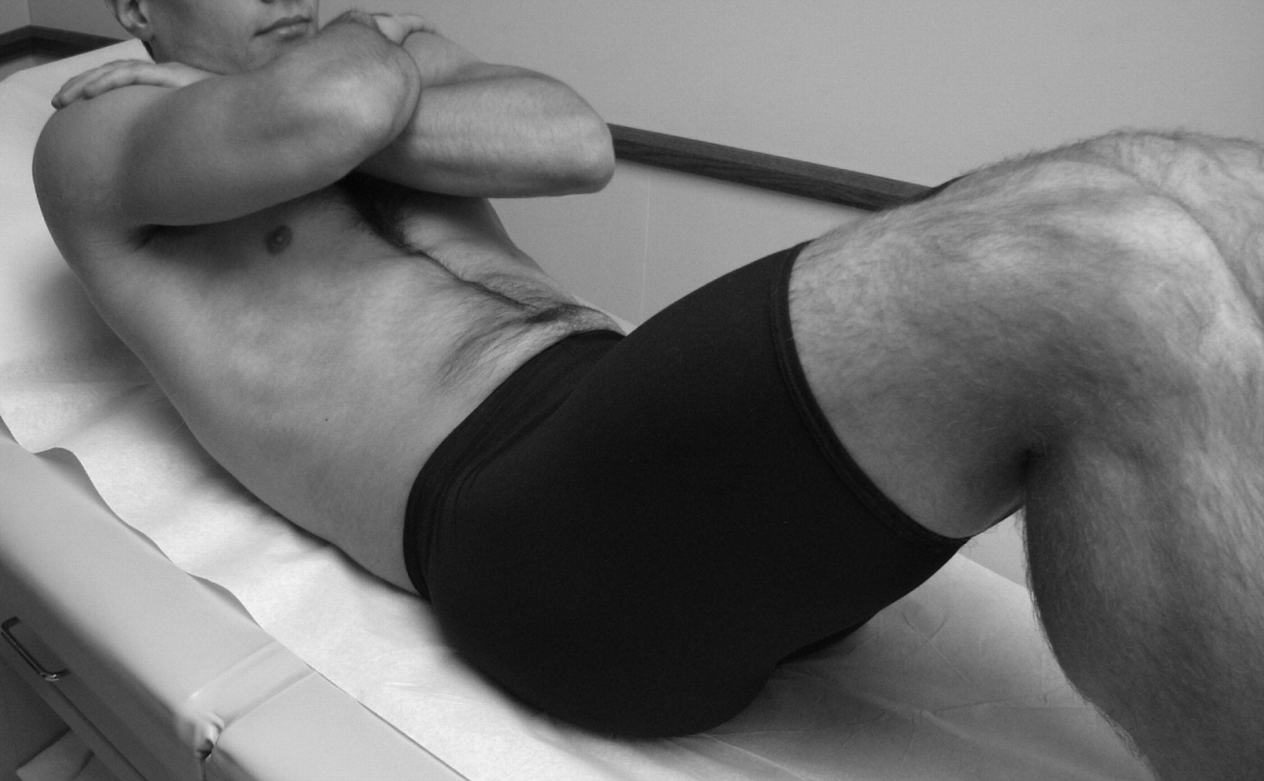
The primary goal when treating quadriceps contusions is firstly to decrease the bleeding into the muscle and secondly to try to minimise knee stiffness. The best way of controlling bleeding and swelling of the thigh is immediately to apply both compression and ice. A specific wrap is indicated for these particular types of injury: the knee is wrapped in a maximally flexed position with a compression wrap and ice (fig 1). This helps not only to decrease the amount of bleeding but also often allows a quicker return to normal knee function and on-ice activities. While the icing helps to decrease the amount of bleeding by vasoconstriction, the compression and wrap helps to make sure that there is less space for the swelling and bleeding to occur. Wrapping the knee in flexion usually allows the athlete to regain their range of motion more quickly. For this particular injury, we have found that, if the knee is iced and wrapped at or near full extension, it usually becomes stiff. It is then hard for the athlete to get back to skating activities until the stiffness is worked out.

Original treatment for a quadriceps contusion. An ice bag is applied over the contusion and a compression wrap is applied to the maximally flexed right knee. This image was staged for demonstration purposes and does not include patient information.
Once a skater’s quadriceps strength and knee motion have returned, they can return to competition. In minor cases, this may be a day or two. In more severe cases, a skater may need to be on crutches for a few days until they can walk without a limp. Athletes who need to cross-train first before they get back to on-ice activities should work on activities that maintain their aerobic and anaerobic endurance. They can jog in a pool or work on a stationary bike as tolerated. In the vast majority of patients, quadriceps contusions will respond very well to a programme of icing, wrapping and bending the knee as much as possible immediately after the injury. In cases in which full motion and strength is not regained within the first few days, further evaluation may be needed.
It is difficult to prophylactically prevent quadriceps contusions. Having the equipment manager check the thigh pads on a team’s defensemen and verifying that the shin pads have sufficient padding over the knee can help to minimise these injuries.
Shoulder separations
AC joint separation is one of the most common ice hockey injuries.1 It usually results from on-ice contact with another player or the boards when a player is checked into the boards. The main types that we see in ice hockey are grades I, II and III.
The treatment and prognosis for return to competition after shoulder separations depends on the grade of the initial injury. The number of weeks it will take for an athlete to return to full competition can almost always be calculated by multiplying the grade of the AC separation by 2. Thus it is usually 1–2 weeks for grade I AC separations, 3–4 weeks for grade II, and 5–7 weeks for grade III.
The initial treatment for AC joint separations is to place ice over the AC joint immediately to minimise bleeding and swelling. We also use a shoulder sling for comfort to support the arm for the first few days after a separation. Our athletes are encouraged to immediately start pendulum exercises and active shoulder motion to try to prevent shoulder stiffness from developing. Once they regain their shoulder range of motion, a rehabilitation programme, focusing on rebuilding the rotator cuff, deltoid and other intrinsic shoulder muscle strength, is performed. Athletes can be allowed to return to on-ice activities once they have full motion and strength of the affected shoulder. In most instances, a protective pad is placed over the AC joint under the shoulder pads to minimise pain and the chance of a recurrent injury, through to the end of the season (fig 2). This protective pad helps to both provide comfort over the injured AC joint and also dissipate contact forces over a wider area of the joint if they do have contact.

A protective acromioclavicular joint sprain pad (right shoulder). This image was staged for demonstration purposes and does not include patient information.
Shoulder separations are the most common shoulder injury in ice hockey. Proper evaluation and treatment can set the guidelines for an expected return to competition and minimise downtime. In general, we recommend that almost all AC separations have x-ray examinations to make sure that there is no associated fracture. The best way to make sure that athletes regain their full strength and motion before returning to play is to verify that they do not have any residual shoulder weakness or stiffness. In addition, an early icing and range of motion programme is essential to minimise downtime.
High ankle sprains
High ankle sprains involve an injury to the distal anterior tibiofibular joint. Whereas the majority of ankle sprains in ice hockey are low ankle sprains and do not require surgery, high ankle sprains can be a real problem for skaters because the blood supply in this area is poor, and it takes a long time for these injuries to heal. Any type of twisting or turning manoeuvre can result in recurrent microtears and irritation of this area, which can delay healing of these injured ligaments. Over time, athletes can have a fairly normal gait pattern, or even jog on level ground normally, but cannot push off or twist on their skate edges, and can be quite limited in terms of their ability to return to transitioning for on-ice activities. High ankle sprains can take up to 6 weeks or longer to heal.
Treatment of these sprains can be very difficult in ice hockey players because they usually cannot return quickly to on-ice activities. While the more common lower ankle sprains can usually be taped and fitted into a tight skate, allowing a reasonably rapid return to skating once the pain and swelling have resolved, it takes much longer for a skater to recover from high ankle sprains. The initial treatment for high ankle sprains is to follow a programme of rest, icing, compression and elevation (RICE). In our experience, an immediate RICE protocol can decrease the downtime for even severe high ankle sprains. If the ankle is allowed to swell after the sprain and the athlete loses ankle motion, return to on-ice activities in a timely fashion is significantly compromised.
Once ankle swelling has decreased and range of motion has improved, the athlete can start a programme of ankle strengthening. We usually use rubber tubing, or have the athlete push against a table leg, to strengthen the peroneal musculature and other ankle muscles to protect the ankle from being reinjured. In addition, it is important to work on an appropriate proprioceptive training programme to allow the ankle to “re-recognise” where the joint is located after injury. Balancing exercises are especially important after high ankle sprains.
Once an athlete has minimal swelling and appropriate strength, a functional on-ice evaluation needs to be performed to determine when they can return to competition. It is especially important in the treatment of high ankle sprains in ice hockey players that they do not try to play through the pain. If players find that they cannot push off on their edges and have difficulty with transitioning from forward to backward skating, the injury still needs to be rested. Trying to skate through the pain will often prolong the total rehabilitation time.
It is much more difficult to treat high ankle sprains than low ankle sprains in ice hockey players because of the constant twisting and turning during on-ice activities. Treatment needs to be individualised to the athlete, and symptoms resulting from twisting and turning need to be resolved before return to skating to minimise downtime.
Sports hernias
Sports hernias in ice hockey often present as a nagging groin strain which will not resolve. Almost by definition, groin strains that linger for more than a few weeks are usually sport-related hernias. Sports hernias are not like inguinal hernias, but rather are injuries to the small and thin abdominal wall muscles attached around the inguinal ring. Because sports hernias have only become recognised in the sports community over the past decade, it is quite common for athletes to see several physicians before the true pathology is identified, or for them to be treated for long periods of time for a suspected groin strain.
The most common complaint of athletes with sports hernias is that they cannot transition for on-ice activities and have difficulty twisting and turning on the affected side. On physical examination, almost all will feel pain on palpation of the injured inguinal ring region, and they also have pain when performing an abdominal crunch or a resisted one-leg straight-leg raise on the affected side (fig 3). In fact, the most common finding on physical examination of patients with a sports hernia is that they have difficulty performing abdominal crunches without pain.
Demonstration of an abdominal crunch. This image was staged for demonstration purposes and does not include patient information.
Unfortunately, as sports hernias involve a tear of the abdominal wall muscles, the only effective treatment is to stop the activities that cause irritation or to have surgery. Surgery is almost always performed by a general or orthopaedic surgeon who specialises in this area. It involves reattaching the abdominal muscles and often placing them in a plastic reinforcing mesh for reinforcement to minimise the chance of tearing again.2 Depending on the surgeon, their experience and their protocol, most athletes return to on-ice activities within 2–4 weeks of surgery.
It is important for an athlete with lingering groin pain to see a sports physician to verify the diagnosis. It is also important to rule out other causes of groin pain such as femoroacetabular impingement (especially in goaltenders), stress fractures, infections and nerve entrapment. Any lingering source of groin strain should be carefully evaluated.
Medial knee injuries
Although the superficial medial collateral ligament, the main medial knee ligament, is one of the strongest in the body, it is the most commonly injured ligament in ice hockey. It is most commonly injured by an on-ice collision causing a valgus stress to the knee. We have also seen it when a skater catches their edge on the ice and sustains a twisting force. We classify medial knee injuries according to the original AMA guidelines of grades I, II and III. Grade I injuries are mild sprains with minimal gapping, grade II are partial tears of the medial knee ligaments, and grade III are complete medial knee injuries with significant medial compartment gapping.3
Treatment of isolated medial knee injuries in ice hockey is based on both previous research and previous results in athletes. As with any associated knee injury, the initial RICE protocol is essential. We immediately treat these injuries with an ice wrap to try to decrease the swelling of the knee joint. We also apply ice for ∼20 min out of each hour for the first 48 h after injury to try to minimise by vasoconstriction bleeding into the surrounding tissues. A compression wrap or elastic sleeve is also applied to attempt to decrease swelling. We also commonly use a hinged knee brace to provide some stability to the healing ligament for grade II and III medial knee tears.
Once the initial phase of treatment is completed and athletes can progress to a rehabilitation programme, they are immediately started on exercising on a stationary bike. Early repetitive cycling motion greatly assists medial knee ligament injuries to heal.4 We start our athletes on a stationary bike as soon as they can tolerate it, and increase the time and resistance according to the amount of swelling of the knee.
Ice hockey players should be followed-up and closely monitored for rapid advancement of this rehabilitation protocol. They may return to competition once they have regained full strength, have no swelling of the knee, and have evidence of healing of the medial knee ligament on physical examination. The general time frame for returning skaters to competition is up to 1–2 weeks for grade I tears, 3–4 weeks for grade II tears, and 4–6 weeks for grade III tears. Athletes who have a complete grade III injury usually return to competition with a hinged knee brace through the end of the season to try to minimise the chance of a recurrent tear.
Lateral (fibular) collateral knee ligament injuries
Injuries to the lateral (fibular) collateral ligament in ice hockey are uncommon because most contact injuries occur in a valgus rather than a varus manoeuvre. However, we do find that these injuries do occur in some athletes. As many ice hockey players are in varus alignment, we find that injuries to the lateral collateral ligament are not tolerated well for on-ice activities and that the athletes are often unstable in side-to-side activities. This is because, when the injury is combined with a varus lower extremity alignment, the lateral knee gapping can be accentuated, making it difficult for any type of push-off or striding activities toward the injured side.
One of the most important things to realise about injuries to the lateral collateral ligament is that, when it is completely torn, it usually does not heal.5 6 Thus it is almost exactly opposite in its natural healing history from medial knee injuries, which almost always heal. Therefore it is important to determine if there is a complete tear of this ligament using a good clinical examination, stress radiographs and MRI scans.7 Complete tears need to be monitored very closely and operated on within the first 2–3 weeks after injury if they do not show evidence of healing.
In cases of partial lateral collateral ligament tears, it is important to give the ligament some time to heal before putting significant stress on it, or it may heal in an elongated position resulting in residual instability problems. We usually recommend that athletes be braced for 2–3 weeks with no significant twisting, turning or pivoting activities allowed before any strenuous activities.
For high level athletes, we recommend the use of a medial compartment unloader brace to allow them to return to competition as soon as possible. We usually have them wear the unloader brace for the first 6–8 weeks after partial tears to protect the ligament from stretching out to become a complete tear. Although it is possible that a well-fitted hinged knee brace may also be effective, we believe that the use of a medial compartment unloader brace significantly minimises the risk of a reinjury.
A good clinical knee examination and varus stress radiographs7 should determine athletes who have partial lateral collateral ligament tears. For athletes who do have partial tears, a standard RICE protocol, an off-ice rehabilitation programme, and the use of a medial compartment unloader brace can assist a more rapid return to competition.
Tears of the anterior cruciate ligament (ACL)
Ice hockey is one of the few sports that can usually be played by an athlete with an ACL tear with minimal chance of injuring the knee further. It is not unreasonable for someone with minimal instability from an ACL tear and no other associated knee injuries to try to rehabilitate their knee and attempt to return to skating to complete the season. However, for skaters found to have significant instability on physical examination or with meniscal tears, we recommend that the ACL be reconstructed to protect the knee before any further intra-articular damage can occur. The issue of whether to use a patellar tendon or hamstring autograft depends on natural laxity, whether other contact sports are played, and what the surgeon does best. In general, a patellar tendon autograft is still considered to be the “gold standard” for an ACL reconstruction in ice hockey players.
In terms of rehabilitation after an ACL reconstruction, for the first few weeks we stress that players should strive to obtain full range of motion of the knee and work on re-activation of the quadriceps mechanism. We have found that athletes who can extend their knee fully immediately after surgery tend to achieve a much quicker return to function. We stress that, for the first couple of weeks after ACL reconstruction surgery, athletes should remain on crutches until they can ambulate without a limp. We also request that they stay in their knee immobiliser until they can perform a straight-leg raise without an extension sag. Once they can do this, we allow them to progress from crutches as tolerated. It is important for the first 6 weeks after surgery that an aggressive biking programme be followed to maintain endurance.
Between weeks 6 and 12, athletes are allowed to increase their activities progressively using a stationary bike, an elliptical machine, a slide board and leg presses as tolerated. Towards the end of this time frame, they may work on more involved exercises and a balancing programme. At 3 months after surgery, most athletes are strong enough to start a jogging and running programme. It is usually at about this time that we allow them to return to skating, but they should not have any contact.
Once athletes have regained full strength and function, which is usually around 5 months after surgery, we test them with KT-1000 and functional testing to determine if they have the appropriate agility, balance and strength to return to full on-ice activities. We have found that athletes who return to competition sooner than this have a higher risk of reinjury of their ACL reconstruction graft, and we generally caution against returning to competition sooner than 4½–5 months after surgery.
Protocol for removing injured players from the ice
Owing to the aggressive nature of ice hockey, it is not uncommon for athletes to have to be removed from the ice on a spine board. Some of the aggressive play is thought to be due to the mandatory use of face guards.8 We have seen on-ice seizures, cervical spine fractures and skull fractures occur in ice hockey players, so it is essential that the team physician understands the correct protocol for removing injured players from the ice. In addition, communication with athletic trainers and emergency medical technicians before games about protocol is essential. The use of the cross-armed sign is recommended as a signal for the team physician and emergency medical technicians to proceed quickly on to the ice to evaluate and possibly transport the athlete.
In the evaluation of an injured athlete, especially one that has been checked from behind or tripped into the boards, it is important to perform the basics of any safety evaluation. The initial first step should always be evaluation of the patient’s airway, breathing and circulation (ABCs).
If the athlete is unconscious, it is important to make sure that they are breathing and also to check their pulse. For an athlete who does not appear to have normal ABCs, the basics of cardiopulmonary resuscitation should be initiated. This is beyond the scope of this article, but every team physician should have training in this. For athletes who are conscious and alert, one of the first things to ask them is where they hurt. Those with any head, neck or back pain should be evaluated very carefully and not moved without assistance. Athletes suffering from loss of breath or extremity pain should be rolled on to their backs so that they can be better evaluated.
One of the most important things to remember when dealing with injured hockey players on the ice, especially when they are unconscious or alert with neck or head pain, is not to remove their helmet. This is because the helmet provides support to the spine, and, if a cervical spine fracture is present, removal of the helmet may cause motion of the fracture, which may lead to permanent nerve damage.9 There is a significant risk of increased motion at the mid-cervical spine level when the helmet is removed, which is the level at which most cervical spine injuries occur in ice hockey.10 Thus it is important to leave the helmet on until it is confirmed that there is no fracture of the cervical spine.
Athletes who have neck pain and are lying in a prone position should not be moved until personnel trained in the correct log rolling technique are available to help move them on to their back.11 At all times, the neck should be stabilised by a physician or the most senior medical person at the scene, and the helmet should be left in place (fig 4). The same protocol should be followed for athletes who have low back pain because they may have a thoracic or lumbar spine injury.

Correct technique for log rolling an injured athlete on the ice to place them on a spine board. This image was staged for demonstration purposes and does not include patient information.
Athletes who are down on the ice but have had a blow to the chest and have simply lost their breath should be allowed to catch their breath before attempts are made to move them. It is also logical to assume that anyone who has hit their head has a concussion until proven otherwise. Athletes who have a headache or neck pain or who are disoriented and appear confused should not be allowed to return to the competition.
To summarise this important topic, any athlete with a potential head or neck injury should not have their helmet removed. For athletes who have isolated extremity pain, splinting of the extremity and assisting them off the ice can be considered. In all cases of head or neck pain, however trivial it may seem, it is important to wait for emergency medical services to help remove the injured player from the ice to make sure that there are no significant cervical spine or other related injuries.
Conclusions
The aggressive nature and speed of skaters in intercollegiate ice hockey commonly results in injuries during competition. Knowledge of common ice hockey injuries and their management is essential in returning athletes to competition quickly. The practical treatment guidelines provided here should serve as baseline knowledge for team physician coverage of this sport.
Acknowledgments
We acknowledge Erik Rasmussen ATC for assistance in obtaining the figures.
Footnotes
Competing interests None.
Provenance and Peer review Not commissioned; externally peer reviewed.