Article Text

Abstract
Background: Although moderate exercise can benefit health, acute and vigorous exercise may have the opposite effect. Strenuous exercise can induce alterations in the physiology and viability of circulating leucocytes, which have a causal relationship with exercise-induced immune distress.
Objectives: To investigate the use of mitochondrial transmembrane potential (MTP), a functional marker of the energy and viability status of leucocytes, for monitoring the immunomodulating effects of short-term, high-intensity exercise.
Methods: 12 healthy volunteers with a mean Vo2max of 70.4 ml/kg/min carried out 3 consecutive days of high-intensity exercise (85% of Vo2max for 30 min every day). Blood samples were collected at multiple time points immediately before and after each exercise session and at 24 and 72 h after the completion of exercise. Leucocyte MTP, apoptosis and circulatory inflammation markers were measured by flow cytometry and enzyme-linked immunosorbent assays.
Results: MTP of peripheral blood leucocytes had declined immediately after the first exercise session and remained subnormal 24 h later. It did not normalise until 72 h after exercise. The sequential changes in MTP were consistent among the three leucocyte subpopulations (polymorphonuclear neutrophils, lymphocytes and monocytes) and were significant (p<0.05). Leucocytes displayed a gradual and incremental change in their propensity for apoptosis during and after exercise. Similarly, plasma concentrations of tumour necrosis factor-α and soluble Fas ligand were raised during the exercise sessions and had not normalised by 72 h after the completion of exercise. Correlation between changes in leucocyte MTP and plasma concentrations of tumour necrosis factor-α and soluble Fas ligand was variable, but significant for polymorphonuclear neutrophils and lymphocytes (p<0.05).
Conclusions: Short-term, high-intensity exercise can lead to a significant and prolonged dysfunction of the mitochondrial energy status of peripheral blood leucocytes, which is accompanied by an increased propensity for apoptosis and raised pro-inflammatory mediators. These results support the immunosuppressive effects of excessive exercise and suggest that MTP is a useful marker of these effects.
Statistics from Altmetric.com
Regular exercise is known to reduce the risk of chronic diseases, such as diabetes,1 cardiovascular disease2 3 and cancer,4 and prevent osteoporosis, obesity and aging. However, the amount of exercise required to achieve beneficial effects has not been clearly defined. To gain maximal health benefits from exercise while avoiding potentially deleterious effects, it is important to determine the “appropriate” amount of physical activity. The frequency, intensity and duration of exercise and pre-exercise fitness level may all be important determinants of the response to exercise.5
Cumulating evidence has shown that intense exercise has adverse effects on different aspects of health. For example, heavy physical activity can trigger an acute myocardial infarction6 and increase the occurrence of premature ventricular depolarisations, which have been associated with a long-term increase in the risk of cardiovascular death.7 A high percentage of apoptosis of lymphocytes has been shown to be induced by intense treadmill exercise, which may in part account for exercise-induced lymphocytopenia and reduced immunity.8 Moreover, it has also been shown that intense exercise induces cellular and circulating oxidative stress and free-radical production.9 10 Our group has shown that leucocyte mitochondrial transmembrane potential (MTP), a marker of the energy and redox status of cells, declined significantly after exhaustive aerobic exercise, and this appeared to have a temporal relationship with intravascular oxidative stress and increased propensity for apoptosis.11
Given the mixed effects of exercise on health, it is important to elucidate the mechanism underlying the different “dose effects” of exercise. In this study, we explored the potential usefulness of leucocyte MTP as a marker of exercise-induced immunosuppression, with emphasis placed on short-term, high-intensity exercise. Our results may help to establish more evidence-based guidelines for the appropriate amount of physical activity and exercise training.
MATERIALS AND METHODS
Subjects
Twelve young trained male runners (mean (SD) age 23.5 (4.9) years, body weight 63.9 (2.4) kg) volunteered to participate in this study. Questionnaires were used to exclude the presence of infectious, inflammatory or immune diseases before the study period. The participants were asked to refrain from any form of training or vigorous physical activity for 2 weeks before the beginning of the study and were not allowed to take any anti-inflammatory agents, steroids, antioxidants or vitamin supplements before, during, or after study entry. The objectives and methods of the study were clearly explained to all of the subjects, who then gave written consent. The research protocol was approved by the Committee for Research on Human Subjects at Taipei Physical Education College and conformed to the guidelines of the Helsinki Conference for Research on Human Subjects.
Exercise model and blood sampling
All subjects underwent a maximal exercise test, using the graded maximal treadmill protocols of Bruce et al.12 Oxygen uptake was measured using a Sensor Medics MMC Horizon System 2900 metabolic cart (Sensor Medics, Anaheim, California, USA). This system uses an autocalibration technique, using 24% O2, 8% CO2 and 12%O2, and 0% CO2 gas tanks. Treadmill speeds corresponding to 85% of maximal oxygen consumption (Vo2max) were calculated for use during the Vo2max exercise test. The mean Vo2max was 70.4 (4.7) ml/kg/min. The participants were then asked to continue their normal daily physical activities without any intense exercise for at least 1 month before the study. During the study, the participants ran on the treadmill at 85% Vo2max for 30 min daily for 3 consecutive days. Venous blood samples (15 ml) were collected from each subject immediately before the first run (D1) and at sequential time points during the exercise period (D1′, immediately after exercise on the first day; D2′, immediately after exercise on the second day; D3′, immediately after exercise on the third day) and then 24 and 72 h after completion of the exercise. A portion of the EDTA-treated blood sample (5 ml) was centrifuged at 1500 g and the plasma was stored at −20°C until subsequent analysis (see below). The remainder of the blood sample was heparinised and assayed immediately for MTP.
Isolation and labelling of leucocytes and measurement of MTP
Measurement of MTP in freshly isolated peripheral blood leucocytes has been previously described.11 Briefly, heparinised blood was treated with an erythrocyte lysing buffer (Harlingen, San Diego, California, USA), washed once with phosphate-buffered saline, and centrifuged at 200 g for 5 min. The cell pellet was resuspended in Hank’s balanced salt solution (Gibco BRL, Paisley, Scotland, UK) to about 105 cells/ml. The resultant leucocyte suspension was then incubated with 5 μmol/l 5,5′,6,6′-tetrachloro-1,1′,3,3′-tetraethylbenzimidazolcarbocyanine iodide (JC-1) (Molecular Probes, Eugene, Oregon, USA) for 20 min at 37°C. After staining, JC-1 was incorporated into the mitochondria, where it formed either monomers (green fluorescence, 527 nm) or, if MTP was high, aggregates (red fluorescence, 590 nm). The ratio between the fluorescence intensity of the JC-1 aggregates and monomers reliably reflects MTP of single cells independently of the change in number and mass of the mitochondria.13 The fluorescence of JC-1 was analysed by cytometry using a FACScan (Becton Dickinson, San Jose, California, USA), and separate electronic gates were set on the dot plot of the forward and side scatter to include the different subpopulations of leucocytes (polymorphonuclear neutrophils (PMNs), monocytes and lymphocytes). To ensure consistency of the data from the different measurements, the photomultiplier value of the detector in the different channels was set at 450 V throughout all the experiments. All the analytical procedures were completed within 3 h of blood sampling.
Assessment of apoptosis
Apoptotic cells were detected by evaluating translocation of membrane phosphatidylserine, an early characteristic of apoptotic cells, with an annexin V assay (BD PharMingen, San Diego, California, USA). Propidium iodide was used as a counterstain for the assay to determine plasma membrane integrity and cell viability. Apoptotic cells were identified as the population of annexin V-positive, propidium iodide-negative cells in the three subpopulations of leucocytes separated by electrical gating as described previously.14 The extent of apoptosis was represented as the percentage of all cells analysed that were apoptotic.
Measurement of plasma tumour necrosis factor (TNF)-α and soluble Fas ligand (sFasL)
Plasma concentrations of TNF-α and sFasL were measured using ultrasensitive ELISA kits (BioSource, Camarill, California, USA).
Statistical analysis
All continuous data were expressed as mean (SEM). Group comparisons were assessed using the Mann-Whitney U test. Sequential changes were assessed by repeated-measures analysis of variance. Pearson’s correlation analysis was used to examine the relationship between variables. p<0.05 was considered significant.
RESULTS
Changes in leucocyte MTP before and after short-term consecutive high-intensity exercise sessions
Compared with before exercise, MTP in all three leucocyte subpopulations had declined significantly immediately after the first exercise session (fig 1). It had then declined more substantially after the second (D2′) and third (D3′) exercise sessions. It remained subnormal up to 24 h after the completion of the 3-day exercise programme and returned to pre-exercise level 72 h after exercise. The sequential alterations in MTP were significant for all three leucocyte subpopulations (p<0.05).

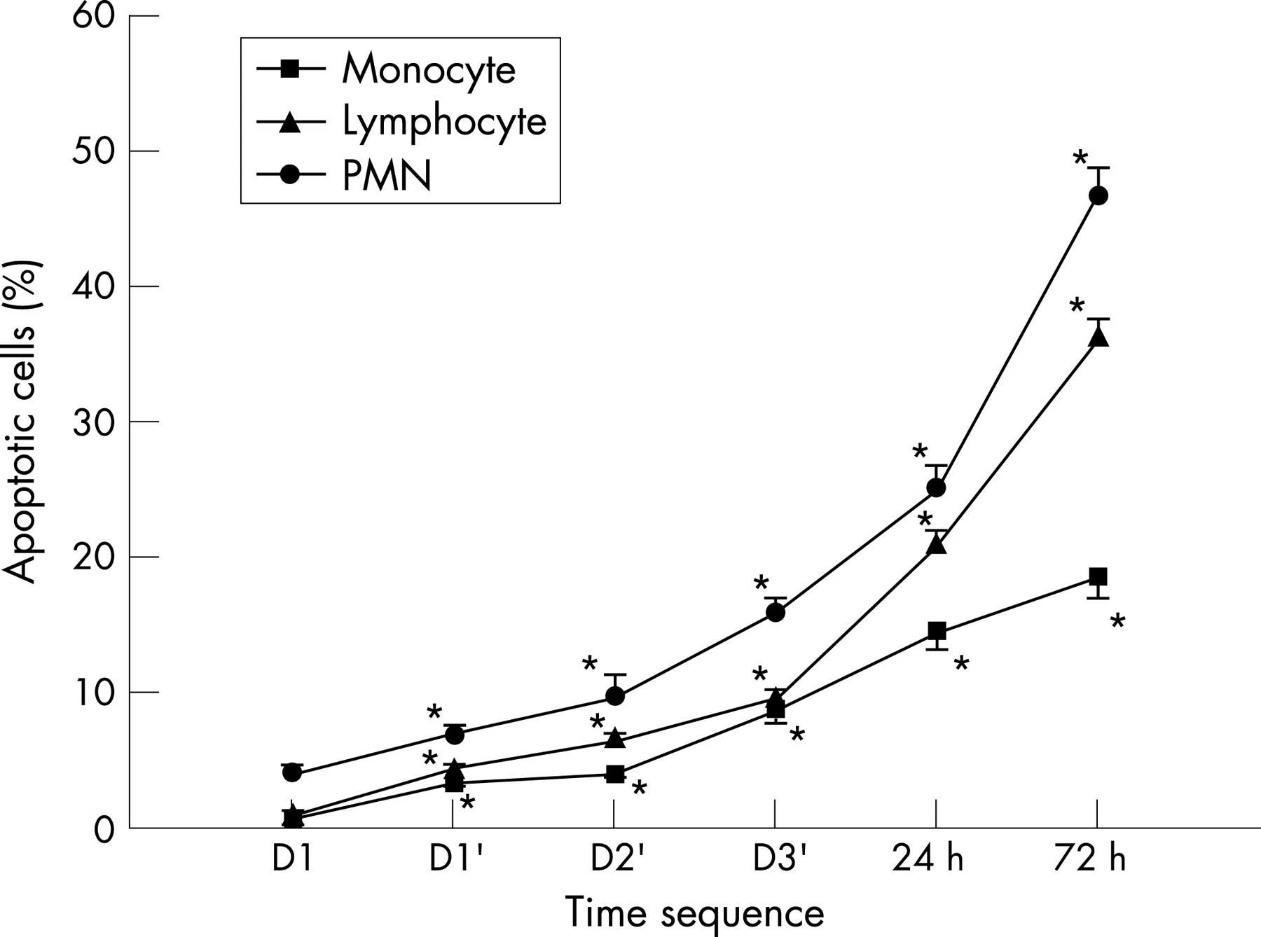
Increased propensity for apoptosis of peripheral leucocytes after short-term consecutive high-intensity exercise sessions
The propensity for apoptosis of PMNs, lymphocytes and monocytes increased gradually after each exercise session (fig 2) and then rapidly after the completion of the 3-day consecutive exercise programme, peaking 72 h after exercise.
Effects of short-term consecutive high-intensity exercise sessions on plasma TNF-α and sFasL concentrations
The plasma concentration of TNF-α had increased significantly (p<0.05) after the first exercise session and remained substantially higher than the pre-exercise level throughout the 3-day exercise programme and until 72 h after exercise (fig 3). Similarly, the plasma concentration of sFasL had increased significantly after the second exercise session and remained abnormally high thereafter. The changes in TNF-α and sFasL were all significant (p<0.05).

Correlation analysis
To examine the correlation between changes in apoptotic regulators, leucocyte mitochondrial alterations and apoptosis, a bivariant correlation of the change in MTP with inflammatory cytokines was evaluated. Changes in MTP in the three subsets of peripheral blood leucocytes correlated variably with the inflammatory cytokines (table 1). Changes in MTP in the lymphocytes correlated positively with peak plasma TNF-α concentration (p<0.05). A significant correlation was also found between the change in MTP of the PMNs and peak plasma sFasL concentration (p<0.05). Peak plasma sFasL concentration correlated significantly with apoptosis of the monocytes after the 3-day exercise programme (p<0.05). Further, the mutual correlation between the change in MTP in the PMNs and apoptosis was also significant after the 3-day exercise programme (p<0.05).
DISCUSSION
Main findings
In this study, we show that plasma concentrations of TNF-α and sFasL were significantly raised after a short-term, high-intensity exercise session. Leucocyte MTP decreased significantly and immediately after each treadmill session, and this was accompanied by a substantial increase in apoptosis of peripheral blood leucocytes. The change in leucocyte MTP correlated variably and significantly with plasma concentrations of TNF-α and sFasL.
Previous studies
The health benefits of regular and moderate exercise have been well documented.1–4 For instance, all-cause mortality is significantly lower in people who expend ⩾2000 kcal a week during exercise.15 The Harvard Alumni Health Study showed a graded inverse relationship between total physical activity and mortality.16 Furthermore, the National Runners’ Health Study showed a continuum of benefits for cardiovascular risk factors with increasing amounts and intensity of physical activity.17 However, it remains unclear as to how much physical activity should be prescribed for the general population.
What is already known on this topic
Although moderate exercise can benefit health, acute and vigorous exercise may induce immune distress by altering the physiology and viability of circulating leucocytes.
What this study adds
Short-term, high-intensity exercise can lead to significant and prolonged dysfunction of the mitochondrial energy status of peripheral blood leucocytes, which is accompanied by an increased propensity for apoptosis and raised concentrations of pro-inflammatory mediators.
In comparison with regular and moderate exercise, several lines of evidence have suggested the potential risk of acute and high-intensity exercise. Exercise can elicit complex changes in the cellular and humoral immune systems, and strenuous exercise may induce inflammatory reactions and immune disturbances.18 For instance, Mars et al8 reported a high percentage of lymphocyte apoptosis after intense treadmill exercise, which might in part account for exercise-induced lymphocytopenia and reduced immunity. Mooren et al19 suggested that endurance exercise such as marathon running could induce apoptosis in lymphocytes. In the present study, the increased expression of pro-apoptotic death receptor ligands indicates that strenuous exercise can augment cell sensitivity to death induction. Consistent with this possibility, heavy exercise has been shown to compromise cell resistance to oxidant-induced apoptosis and increase active caspase-8, caspase-9 and caspase-3 content of lymphocytes.20 In addition to immune disturbances, the energy needed for the increased metabolic demand during heavy and prolonged exercise is produced by oxidative metabolism, which may overwhelm endogenous antioxidative capacity and cause oxidative damage to cells and tissue.9 10 21 A well-known example of exercise-induced oxidative damage is DNA damage of leucocytes induced by high-intensity aerobic exercise.22–26 According to these studies, strenuous exercise can induce the formation of reactive oxygen species, causing oxidative stress in affected tissues or body compartments.21 The accentuated production of reactive oxygen species may induce increased expression of death receptors and ligands,27 as well as disruption of leucocyte MTP.28 The resultant mitochondrial depolarisation may initiate cellular apoptosis by depleting the cells of their ATP or by releasing pro-apoptotic molecules, such as cytochrome c or apoptosis-inducing factor.28
The variable correlation between the change in leucocyte MTP and apoptotic regulators implies that the leucocyte mitochondrial alterations are part of the systemic immune disturbance induced by short-term, high-intensity exercise. The increased inflammatory cytokines and apoptotic regulators observed after exhaustive exercise may have deleterious effects on peripheral blood leucocytes and may result in changes in MTP. However, the exact causal relationship between them remains elusive. Further studies at a molecular level are required to elucidate the detailed mechanisms underlying these changes. In this study, the delayed appearance of leucocyte apoptosis after mitochondrial depolarisation probably reflects the temporal effect of the apoptotic programme,29 as mitochondrial alterations are known to be early events of apoptosis and to precede other hallmarks of cell death.30 Alternatively, it may reflect the delayed clearance of the apoptotic-cell population from the circulation. Further studies are required to investigate these possibilities.
Study limitations
There are several limitations to this study. Firstly, the subjects were well-trained male runners. Whether the results can be applied to ordinary people and/or female subjects remains unclear. Secondly, the study did not include a control group. However, it is unlikely that healthy athletes would display significant alterations in leucocyte MTP and immune function without performing high-intensity exercise. Thirdly, because of the limited sample amounts, other important inflammatory markers, such as interleukin-6 and C reactive protein, were not examined.
CONCLUSION
We show in this study that short-term, high-intensity exercise can lead to significant dysfunction of the mitochondrial energy status in peripheral blood immune cells, which is accompanied by an increased propensity for apoptosis and an increase in pro-apoptotic cytokines. The results support the potentially deleterious effects of excessive exercise on immune function and health.
Acknowledgments
We thank Ms Pei-Feng Wu for her excellent technical assistance.
REFERENCES
Footnotes
Competing interests: None.
Linked Articles
- Warm up