Article Text

Abstract
Introduction Several painful conditions throughout the human body—such as interstitial cystitis, Dupuytren's contracture, and complex regional pain syndrome—have been shown to exhibit tissues with an increase in mast cell population. One such painful condition—where the pain is not considered proportionate to the tissue changes—is tendinopathy. One study has shown that activation via protease-activated receptor 4 (PAR4) can lead to an increased nociception in joints, and similar mechanisms may be at play in tendinopathy, where substances released by mast cells may activate the PARs. Mast cells are also known mediators of inflammatory responses to injury, but have also been found to regulate cell proliferation and vasodilation in certain tissues via PARs, and tendinopathies are known to exhibit tissue characteristics involving hypercellularity and vascular changes.
The aim of this study was to determine whether tissues surrounding the Achilles tendon exhibit PAR4, as a basis for further studies on interactions between the tendon, vascularity and nerves, and the mast cell population found in tendinopathy tendons.
Methods Biopsies of about 5×5 mm were taken from the ventral part of the Achilles tendon and the paratendinous tissues of this area, from two groups. One group consisted of patients suffering from mid-portion tendinopathy with tendinosis (n=8). Normal controls (n=2) donated biopsies from the same area. Both procedures were guided by ultrasound and performed through a minimal skin incision. These biopsies were later sectioned and immunocytochemistry was used to study the expression patterns of PAR4.
Double-staining with PAR4 and substance P (SP) was performed.
Results The sections showed strong reactivity for PAR4 in the blood vessels both in the tendon tissue proper as well as in the more abundant vascular areas ventral to the tendon in biopsies from both normal tendons and those from patients diagnosed with tendinosis. Tenocytes expressed PAR4 to a varying degree (figure 1), with it being expressed more in cells with a rounded nuclei, primarily in tendons from the tendinosis group. PAR4 was expressed on SP-positive nerve fibres in the paratendinous tissue (figure 2).
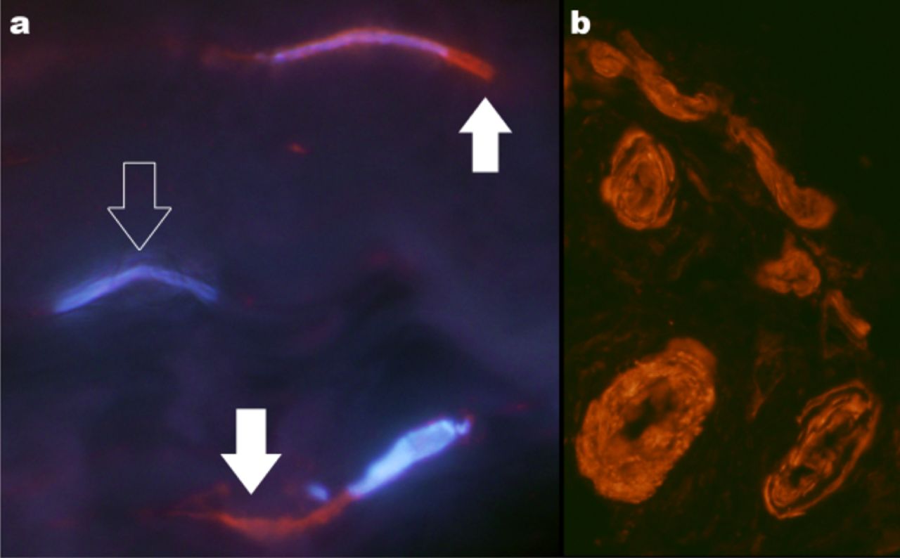
(a) Tenocytes show reactions for PAR4 (in red). Counterstained with DAPI. (b) Vessels positive for PAR4.

(a) A nerve fibre and vessel positive for PAR4. In b the nerve fibre shows reactivity for substance P.
Discussion These findings give basis for further studies into the interactions of SP-positive nerves and mast cells in tendon pathology. Earlier studies have shown regulation of vascular permeability and dilation, as well as fibroblast proliferation through activation of PARs. The PAR4 may, through activation of proteases released from mast cells, to some extent be responsible not only for tenocyte proliferation and vascular regulation, but also for an enhanced pain signalling in tendinopathy through SP-positive afferents.