Article Text

Abstract
Introduction Substance P (SP) and its preferred receptor, the neurokinin-1 receptor (NK-1 R), are expressed in human tenocytes and this is particularly seen in the tenocytes in cases of tendinosis. Hypercellularity, angiogenesis, and collagen disorganisation are common tissue features of this disease. Excessive apoptosis is also microscopically observed in tendinosis tissues. The role of SP and NK-1 R in the regulation of apoptosis and cell survival of tenocytes is poorly understood, but we have previously shown that SP increases tenocyte hypercellularity both in vitro and in vivo. It is possible that SP contributes to tenocyte hypercellularity by both stimulation of proliferation and inhibition of apoptosis.
Akt, a protein kinase also called protein kinase B and known to be phosphorylated into its active form by SP, plays a critical role in controlling the balance of cell survival and apoptosis in other cell types. Activated/phosphorylated Akt (Phos-Akt) promotes cell survival and inhibits apoptosis by targeting the pro-apoptotic Bcl-2 family (which otherwise cause cytochrome C leakage from the mitochondria), and also by regulating expression of anti-apoptotic Bcl-2 family members and caspases. Furthermore, Akt activation is known to protect cells against apoptosis agents belonging to the TNF family of death ligands, such as Fas ligand (FasL) which acts through the Fas receptor (FasR).
The focus of this study is to investigate (1) if FasR stimulation (Anti-Fas) is a good apoptosis model for human tenocytes, (2) if SP protects from Anti-Fas induced apoptosis, and (3) by which mechanisms SP mediates a possible anti-apoptotic response.
Methods Human Achilles tenocytes were grown as primary cultures and used for experiment at passage 3–5. All experiments were performed in serum-starved conditions. Cell viability (crystal violet), TUNEL-staining, and cytotoxicity assay (measuring lactate dehydrogenase (LDH)) were used to evaluate the endpoint effect of Anti-Fas alone or together with SP and/or a specific NK-1 R inhibitor. Immunocytochemistry, qPCR, and Western Blot were used to determine the pathways of Anti-Fas induced apoptosis and the specific inhibitory effect of SP.
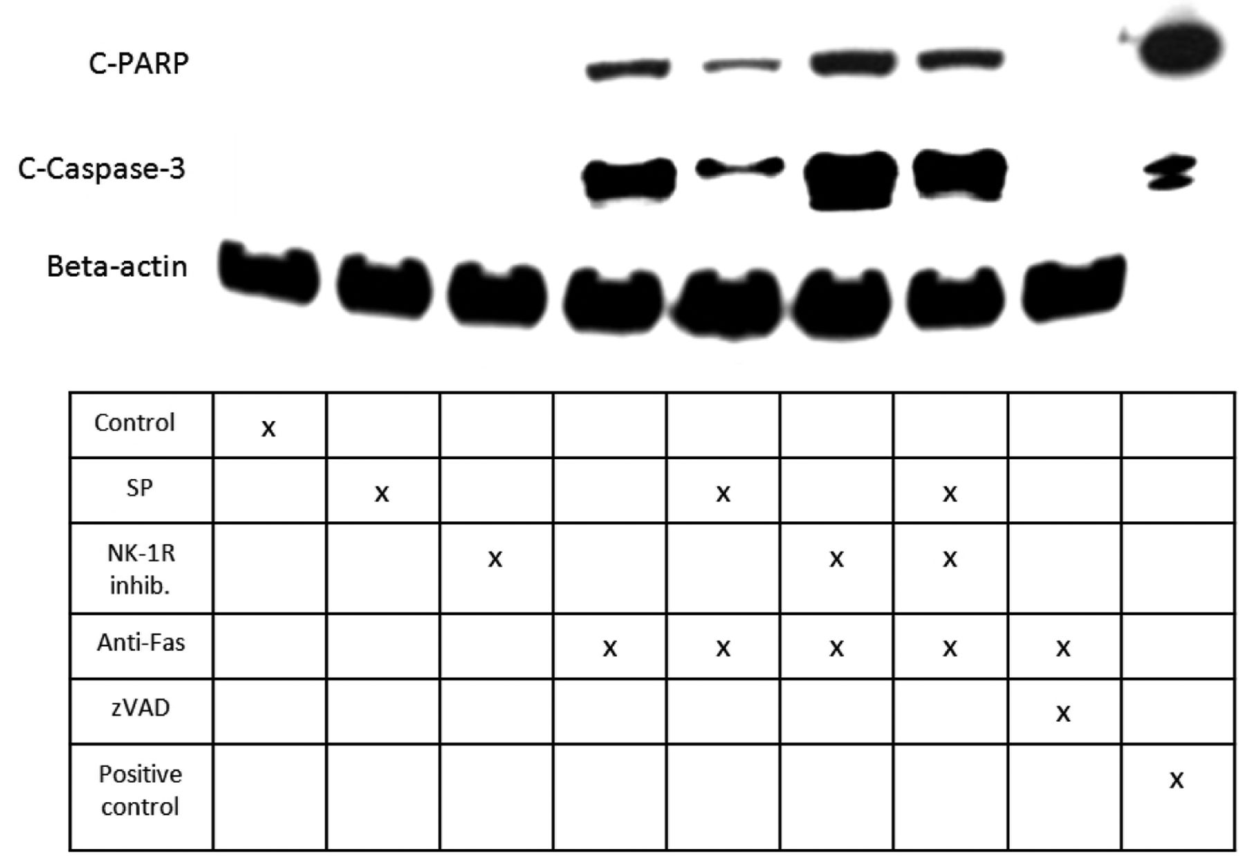
Results The majority of the tenocytes expressed FasR. The LDH assay demonstrated that Anti-Fas treatment results in a time- and dose-dependent release of LDH, and that SP dose-dependently reduces the Anti-Fas induced release of LDH. In parallel, the same trend was seen for the TUNEL assay, that is, SP reduced Anti-Fas induced apoptosis via a NK-1 R specific pathway. In addition it was shown that SP reduced the Anti-Fas induced decrease in cell viability. mRNA and/or protein analysis confirmed that the Anti-Fas induced activation of caspase-8, caspase-3, BID, BAX, and PARP were all down-regulated when SP was included, and that this SP effect was mediated through a NK-1 R specific pathway (for PARP and caspase-3, see figure 1). SP treatment resulted in activation of Akt, and by inhibiting Akt the anti-apoptotic effect of SP was confirmed to be, at least partly, induced through an Akt-dependent pathway.

Cleavage of PARP and caspase-3 after Anti-Fas treatment alone or together with SP and/or the NK-1 R inhibitor illustrating the anti-apoptotic effect of SP. Pan-caspase inhibitor zVAD confirms a caspase dependent cleavage of PARP.
Discussion We show that SP reduces Anti-Fas induced apoptosis in human tenocytes and that this anti-apoptotic effect of SP is mediated through the NK-1 R and an Akt pathway. Considering previous results that SP has a proliferative effect on tenocytes, the present study identifies SP as a potent regulator of cell-turnover in tendinosis tissue, capable of stimulating hypercellularity through different mechanisms. The fact that the FasR is abundantly expressed in tenocytes and that a possible source of the Fas ligand in tendinosis is the inflammatory infiltrated cells in the paratendon, makes it possible that the excessive apoptosis seen in tendinosis is partly explained by FasR stimulation. Most interestingly, as tenocytes are known to endogenously produce SP and express the NK-1 R, it is possible that SP in an autocrine loop rescues cells from Anti-Fas induced apoptosis, via Akt stimulation, thus contributing to tenocyte hypercellularity in tendinosis.