Article Text

Abstract
Introduction The presence or absence of inflammatory cells in chronic Achilles tendinopathy has been a controversial subject in previous studies. Macrophages, T lymphocytes, and neutrophils have previously been detected in tendinopathic human Achilles tendons, whereas other authors have reported that there is no evidence for their occurrence. This controversy may stem from the fact that human Achilles tendon overuse injuries usually develop gradually over time, and the time course of inflammation in response to overuse has been difficult to establish in clinical populations. The aim of this study was to examine the presence of inflammatory cells in the Achilles tendon of rabbits that were subjected to repetitive mechanical loading of defined durations.
Methods In this study, tissue blocks of Achilles tendon taken from a cohort of rabbits on which we previously reported1 were used to analyse the presence of inflammatory cells. Twenty-Four New Zealand male rabbits were subjected to repetitive mechanical loading of the Achilles tendon and grouped into four groups, according to the exercise time period: 0, 1, 3, and 6 weeks. Achilles tendons were harvested at the end of each time period. T-lymphyocytes and neutrophils were examined using immunohistochemistry, and macrophages were identified with Prussian blue staining. All areas of positively labelled cells were captured in digital micrographs using a 20x objective lens and expressed as cell density / viewing field.
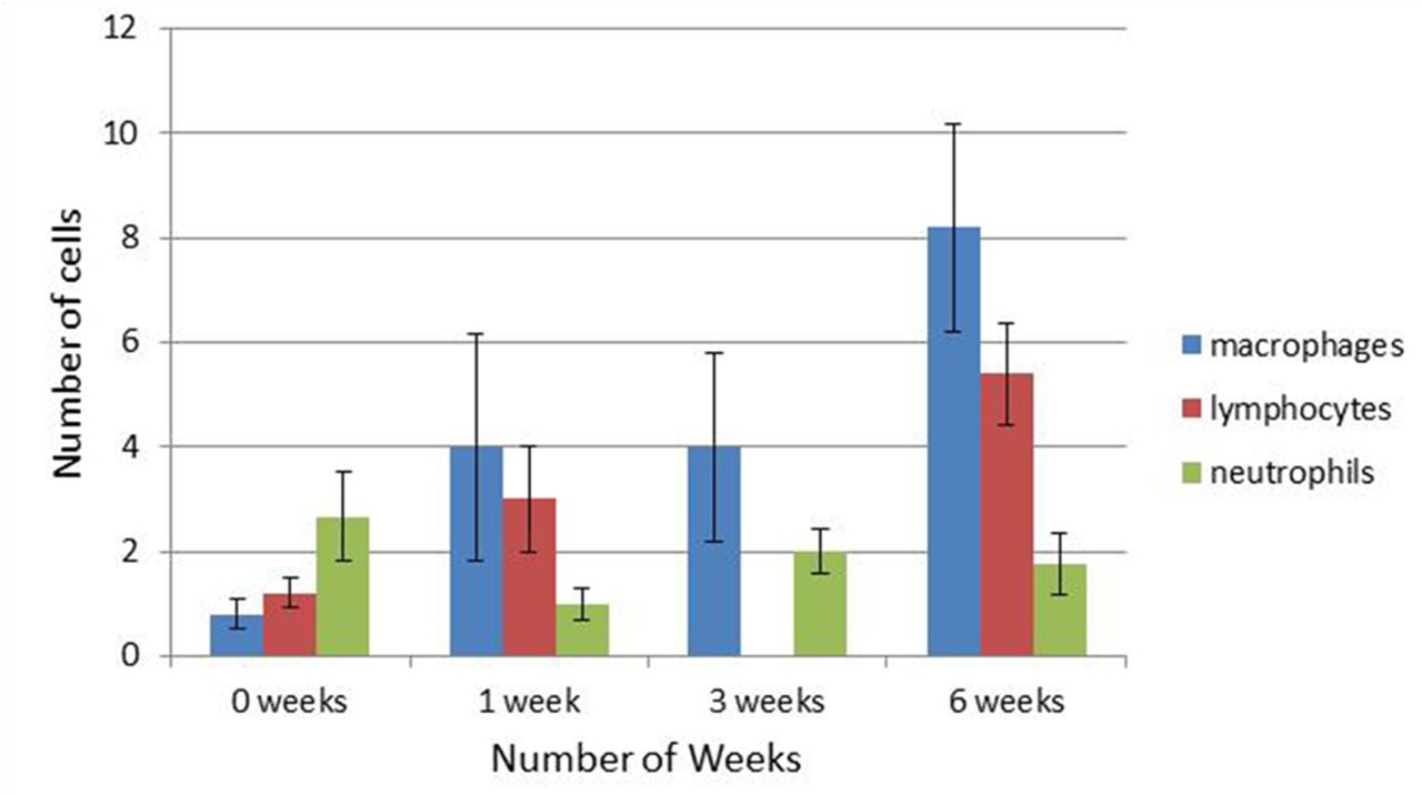
Results Macrophages, T-lymphocytes, and neutrophils were detected in tendon sections from groups 0, 1, 3, 6 weeks. While there was no apparent change in the density of neutrophils over the 6 week time course, the density of lymphocytes (n.s.) and macrophages (p < 0.05) increased over the 6 weeks of overuse (Figure 1). Qualitatively speaking, the evidence of inflammation was not evenly distributed, as some tissue sections from the same groups showed no evidence of inflammatory cells. Inflammatory cells were observed primarily in the paratendon rather than the tendon proper.
Evidence of inflammatory cell involvement during periods of Achilles tendon overuse
Discussion An increasing number of macrophages and lymphocytes were detected in the Achilles tendons of animals subjected to repetitive mechanical loading, with an absence of the same types of cells in some sections from the same groups for unknown reasons. Future studies are needed to (1) confirm these findings using a corroborating technique, e.g. qPCR, and (2) examine the influence of additional variables including tendon region and sex.2 Future studies could examine whether inhibiting inflammation would lessen the extent of tendinopathy in this overuse model.
References Andersson, et al. Br J Sports Med. 2011;45(13):1017–2
Huisman, et al. J Anat. 2014;224(5):538–47