Article Text

Abstract
Context Olympic athletes represent model of success in our society, by enduring strenuous conditioning programmes and achieving astonishing performances. They also raise scientific and clinical interest, with regard to medical care and prevalence of cardiovascular (CV) abnormalities.
Objective Our aim was to assess the prevalence and type of CV abnormalities in this selected athlete's cohort.
Design, setting and participants 2352 Olympic athletes, mean age 25±6, 64% men, competing in 31 summer or 15 winter sports, were examined with history, physical examination, 12-lead and exercise ECG and echocardiography. Additional testing (cardiac MRI, CT scan) or electrophysiological assessments were selectively performed when indicated.
Main outcome measures Prevalence and type of CV findings, abnormalities and diseases found in Olympic athletes over 10 years.
Results A subset of 92 athletes (3.9%) showed abnormal CV findings. Structural abnormalities included inherited cardiomyopathies (n=4), coronary artery disease (n=1), perimyocarditis (n=4), myocardial bridges (n=2), valvular and congenital diseases (n=45) and systemic hypertension (n=10). Primary electrical diseases included atrial fibrillation (n=2), supraventricular reciprocating tachycardia (n=14), complex ventricular tachyarrhythmias (non-sustained ventricular tachycardia, n=7; bidirectional ventricular tachycardia, n=1) or major conduction disorders (Wolff-Parkinson-White (WPW), n=1; Long QT syndrome (LQTS), n=2).
Conclusions Our study revealed an unexpected prevalence of CV abnormalities among Olympic athletes, including a small, but not negligible proportion of pathological conditions at risk. This observation suggests that Olympic athletes, despite the absence of symptoms or astonishing performances, are not immune from CV disorders and might be exposed to unforeseen high-risk during sport activity.
- Evaluation
- Athlete's heart
- Olympics
- Prevention
- Heart disease
Statistics from Altmetric.com
Introduction
Olympic athletes represent a unique subset of the athletic population by virtue of their motivation to excel, strenuous exercise conditioning and astonishing performances. Participating and succeeding in Olympic Games represents the highest achievement in the athlete's career, and the way to gather large visibility, social fame and economic success. It is a fact that Olympic athletes draw special coverage by the media and the lay public. As a mere example, 3.6 billion TV viewers followed worldwide the previous 2012 London Games.1
Olympic athletes also raise scientific and clinical interest, specifically regarding the criteria for eligibility and the most appropriate medical care.2 The rare, but dramatic occurrence of sudden cardiac death/cardiac arrest (SCD/CA) in elite competitors has, over the last decade, further emphasised this issue.3–5
In the present study we sought, therefore, to assess the prevalence and type of cardiovascular (CV) abnormalities in a large cohort of Olympic athletes. To the scope, we took advantage of the data set derived from Italian Olympic athletes, evaluated for CV diseases in the context of a more comprehensive medical programme.6
Methods
Study population
For the present study, we considered athletes evaluated in a 10-year period (from the Olympic Games in Athens 2004 to Sochi 2014) within our CV programme, which included medical history, physical examination, 12-lead and exercise ECG, and echocardiography.7 The Olympic medical programme is implemented in our institution (and funded by the Italian Olympic Committee) for athletes selected to participate to the Olympic Games. Among the 2389 athletes entering the programme, 37 had either incomplete or inadequate data and were excluded. The final study population comprised, therefore, 2352 Italian Olympic athletes.
Athletes aged 14–46 years (mean 25±6), with 64% males, competing in 31 summer or 15 winter sport disciplines were recruited. We arbitrarily classified sport activities in four groups,8 that is, (1) skill (such as table tennis, equestrian, shooting, yachting, curling; n=572); (2) power (weightlifting, sprinting, downhill skiing; n=414); (3) mixed (soccer, basketball, volleyball, handball, water polo, fencing, tennis; n=664) and (4) endurance disciplines (rowing, long-distance running and marathon, cycling, triathlon, pentathlon, cross-country skiing; n=702).
Athletes have been involved in training programmes for 3–12 years (average 6) and continued to train intensely at the time of the study. All were world-class competitors, and a substantial proportion (45%) had participated in more than one Olympic Game.
Written informed consent was waived for athletes undergoing a standard evaluation pursuant to Italian law and the Institute policy. The study design was approved by the Review Board of the Institute and funded by the Italian National Olympic Committee. All clinical data assembled from athletes are maintained in an institutional database.
CV evaluation
Physical examination with personal and family history was performed according to the recommendations of the IOC.2 Blood pressure was measured according to the European Society of Hypertension (ESH)/European Society of Cardiology (ESC) guidelines.9
The 12-lead ECG was recorded with the participant in supine position during quiet respiration, at 25 mm/s, using a Cardioline ClickECG, (Cardioline, Italy). ECG analysis was performed according to criteria distinguishing normal versus abnormal ECG patterns in athletes, as previously reported.10–12
The exercise testing was performed on bicycle ergometer (Cardioline XR400, Cardioline, Italy) with an incremental protocol until exhaustion. ECG was continuously monitored during exercise and 7 min of recovery.
Two-dimensional and Doppler echocardiography was performed by expert cardiologists using an iE33 ultrasound machine (Philips Medical System, Andover, Massachusetts, USA). Two-dimensional measurements of cardiac structures, left ventricle (LV) ejection fraction and mass were performed as recommended by the American Society of Echocardiography/European Association of Cardiovascular Imaging.13 LV inflow velocities and tissue Doppler imaging signals were recorded in the apical four chamber, as previously described.14
Additional investigations
Detection of >1 premature ventricular beat (PVB) on the 12-lead ECG and/or ≥3 PVBs during exercise testing, and/or history of palpitation was indication for 24-hour ECG monitoring.15
Additional tests were selectively performed to confirm diagnosis and/or assess risk stratification in athletes with abnormal ECGs (most commonly, diffusely flat or inverted T-wave), ventricular tachyarrhythmia, or borderline echocardiographic findings (ie, grey-zone of LV hypertrophy), and included CV MR (CMR, n=36), CT scan (n=3), coronary angiography (n=2). Electrophysiological study (n=28), electroanatomic mapping (CARTO, n=5) and radiofrequency ablation (RFA, n=15) were selectively performed in athletes with either sustained supraventricular or complex ventricular tachyarrhythmias.
Statistical analysis
The study design was a retrospective analysis of the CV abnormalities prevalence among Italian Olympic athletes. Continuous data are expressed as mean±SD. Gender differences were evaluated by unpaired-samples t-test. Differences between sport disciplines were evaluated using analysis of variance. Differences between proportions were evaluated by χ2 test. Statistical analysis was performed using SPSS (V.22, SPSS, Chicago, Illinois, USA).
Results
Demographic and CV characteristics
Mean age was 26±5 years for men and 23±4 for women (p<0.001). Men had weight, height and body surface area larger than women. Systolic and diastolic blood pressures were relatively higher, while heart rate was lower in men. Cardiac dimensions (LV cavity, wall thickness and mass) were larger in men (table 1).
Demographic and cardiovascular characteristics of 2352 Olympic athletes according to gender
When analysed by type of sport, athletes competing in mixed disciplines showed the largest body size. Endurance athletes showed the lowest heart rate and relatively high blood pressure. Cardiac dimensions (LV cavity, wall thickness and mass) were larger in endurance compared with remaining athletes (table 2).
Demographic and cardiovascular characteristics of 2352 Olympic athletes according to type of sport participated
Clinical findings
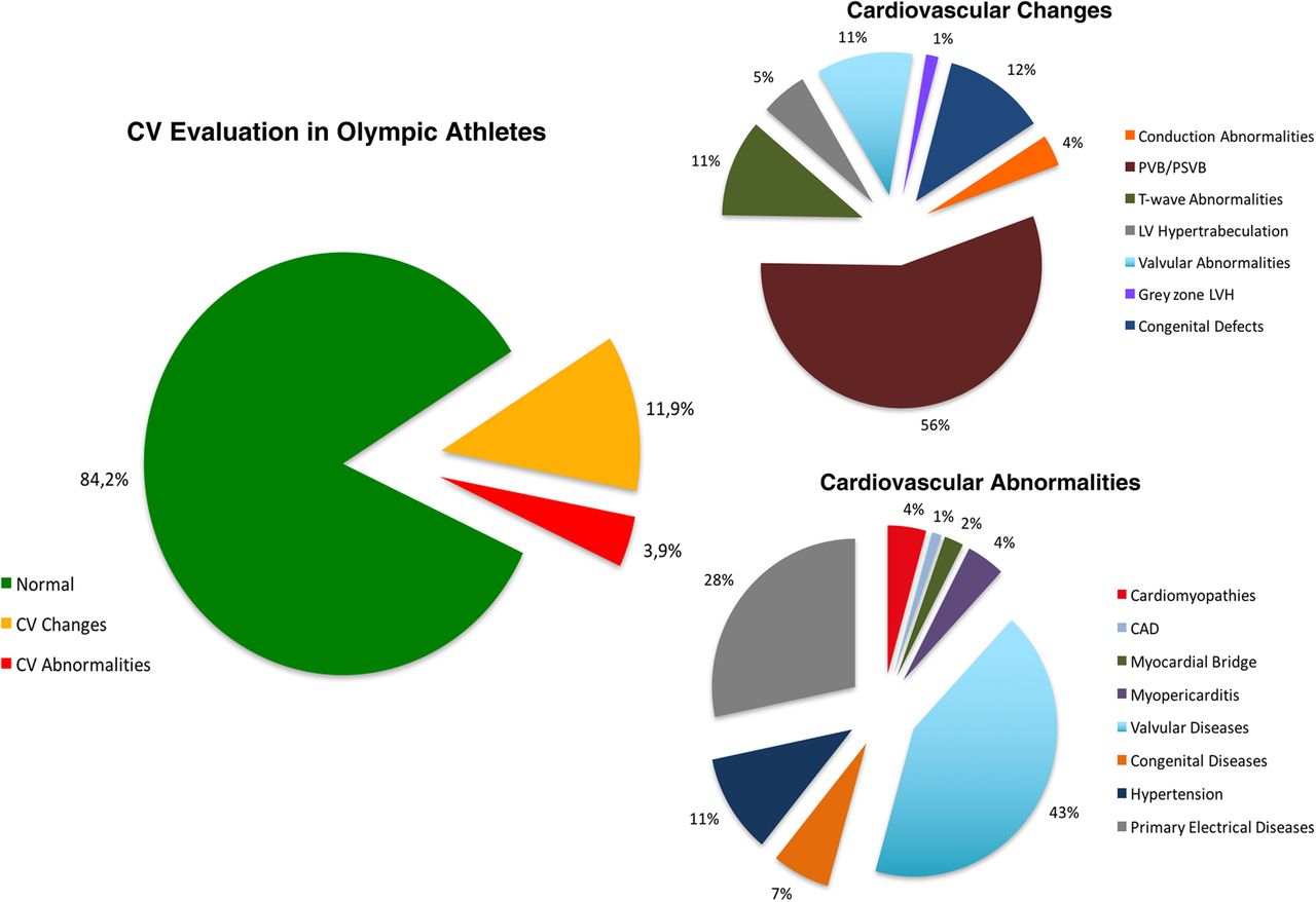
Vast majority of the 2352 athletes, that is, 1979 (84.2%), presented completely normal CV findings. A substantial subset of 281 athletes (11.9%) showed ECG or echocardiographic changes (mostly, isolated PVBs, flat/inverted T-wave, LV hypertrabeculation, see figure 1), which after thorough investigation were considered not to represent pathological cardiac conditions, and deprived of clinical relevance.

The results of CV evaluation in Olympic athletes. Cardiovascular abnormalities: see detailed explanation in the manuscript. Minor cardiovascular changes: conduction abnormalities included 6 athletes with right bundle branch block, 2 with left anterior fascicular block, 2 with bi-fascicular block (n=10). PVB/premature supraventricular beats (PSVB), isolated and rare premature ventricular beats/supraventricular beats in the absence of structural cardiac abnormality at the imaging testing (n=157); T-wave abnormalities, either inverted or diffusely flat T-wave in the absence of structural CV abnormalities on imaging testing (n=31); LV hypertrabeculation, prominent LV trabecular pattern, in the absence of LV dysfunction, abnormal clinical findings and positive family history, judged not to meet the criteria for left ventricular non-compaction (LVNC) (n=15); valvular changes included mild elongation of the mitral valves without evidence of prolapse (n=25), mild asymmetric tricuspid aortic valve without raphe (n=6); grey zone of LVH=LV wall thickness of 13–15 mm, associated with increased cavity size and normal diastolic function, compatible with sport-induced physiological LV remodelling (athlete's heart, n=4). Congenital changes included atrial septal aneurysm (n=13), patent foramen oval (n=9) and corrected atrial septal defects (n=11). CAD, coronary artery disease; CV, cardiovascular; LV, left ventricle; LVH, LV hypertrophy; PSVB, premature supraventricular beats; PVB, premature ventricular beat.
The remaining 92 athletes (3.9%) presented CV abnormalities, judged to be clinically relevant, including 66 (2.8%) with structural abnormalities and 26 (1.1%) with primary electrical diseases (figure 1).
Structural CV abnormalities
Cardiomyopathies were identified in four asymptomatic athletes. Specifically, arrhythmogenic right ventricular cardiomyopathy (ARVC)16 was diagnosed in a cyclist with PVBs with left bundle branch block (LBBB) morphology on exercise-ECG and 24-hour ECG monitoring. RV wall thinning, aneurysm and deranged trabecular pattern were evident on imaging testing. The electroanatomic mapping (CARTO) showed low voltages in the thinned area of right ventricular (RV) free wall. In another pentathlon athlete, diagnosis of ARVC was prompted by the deeply inverted T-waves in anterior precordial leads (V1-V4) on 12-lead ECG, and non-sustained ventricular tachycardia (NSVT) with LBBB morphology and superior axis on 24-hour ECG monitoring. CMR and echocardiography showed only mild RV enlargement. Family screening revealed ECG repolarisation abnormalities and ventricular tachyarrhythmias in the brother.
Hypertrophic cardiomyopathy (HCM)17 ,18 was identified in a swimmer, showing inverted T-wave in aVL, biphasic in V2, V3 on 12-lead ECG. Both NSVT and frequent PVBs were present at 24-hour ECG monitoring. Imaging testing showed an asymmetric LV hypertrophy (maximum thickness of 15–16 mm in anterior septum) and a non-dilated LV cavity (LV diameter, 49 mm). A causative mutation in the MyBPC-3 gene confirmed the diagnosis. In a water-polo player suspicion for HCM was prompted by the asymmetric LV hypertrophy (anterior and posterior septum, 16 mm; posterior free wall, 10 mm), which did not regress after detraining, and was associated with a small LV cavity (diastolic diameter, 48 mm). ECG showed small Q-wave in inferior and lateral leads.
Coronary artery disease was diagnosed in a 33-year-old male rower, presenting a 2 mm ST/T depression on exercise ECG, associated with NSVT and polymorphic PVBs. Subsequent coronary CT scan revealed a soft plaque, with 50% stenosis, on the proximal left anterior descending (LAD) coronary. The athlete had hypercholesterolaemia (low-density lipoprotein, 147 mg/dL, or 3.8 mmol/L) and positive family history for ischaemic heart disease.
Clinical diagnosis of perimyocarditis was prompted in two athletes by the evidence of preceding or concomitant upper respiratory condition, associated with positive inflammatory markers (elevated C-reactive protein) and positive imaging testing.19 Echocardiography showed mild pericardial effusion (in both), associated with apical wall motion anomalies (in one). Two additional athletes with previous and recent diagnosis of perimyocarditis showed persistent mild pericardial effusion and ventricular arrhythmias (PVBs and/or NSVT). Both these findings, in the context of the clinical and laboratory resolution of the inflammatory process, were interpreted as residual and possibly permanent signs of the healing process.
Myocardial bridge was identified in two athletes. A 34-year-old free-style skier with 2 mm ST segment depression on exercise ECG, showed on sestamibi scintigraphy a reversible perfusion defect of the anterior LV wall. Coronary angiography demonstrated a 2 mm deep and lengthy intramural tract of the distal LAD. Another 30-year-old marathon runner presented 3 mm ST segment depression on exercise ECG. CT scan revealed a short and 1.5 mm deep intramyocardial course of the proximal LAD; myocardial scintigraphy failed to show any perfusion impairment.
Vast majority of structural abnormalities comprised either mitral valve prolapse (n=24) or bicuspid aortic valve (n=10), here defined according to the American Heart Association (AHA) task force criteria.20 ,21 Both conditions were associated in one. Less frequent lesions included mild rheumatic aortic stenosis (n=1), mild (n=2) or severe (n=1) pulmonary stenosis, atrial septal defect (n=4) and patent ductus arteriosus (n=2).
Finally, systemic hypertension was diagnosed in 10 athletes by blood pressure values consistently ≥140 and/or 90 mm Hg on repeated measurements.9 Of them, six reported positive family history for hypertension.
Primary electrical diseases
Primary electrical diseases comprised supraventricular or ventricular tachyarrhythmias (n=23), or major conduction defects (n=3).22 Recurrent episodes of paroxysmal atrial fibrillation (AF) with rapid ventricular rate (up to 200 bpm) were recorded on 24-hour ECG monitoring and exercise ECG in a female tennis player, successfully resolved by RFA. Brief, repeated short episodes of AF with low ventricular rate (average 70 bpm) were recorded on ECG in a young triathlon athlete. In 14 other athletes, all reporting of palpitations, prolonged runs of supraventricular reciprocating tachycardia (SVT) were identified by repeated 24-hour ECGs monitoring.
Bursts of NSVT (3–16 beats) were detected on exercise and/or 24-hour ECG monitoring in six athletes (three of them reported palpitations), associated with frequent and/or polymorphic PVBs (in three). Another athlete showed exercise-induced frequent, polymorphic PVBs and short bidirectional NSVT, suggestive for catecholaminergic ventricular tachycardia (VT).
A Wolff-Parkinson-White pattern was identified in a baseball player. A long QT interval was suspected in a female judo player, with QTc of 0.48 s, with negative gene analysis. LQT1was confirmed by presence of mutation in KCNQ1 in an open-water male swimmer, with QTc interval of 0.47 s, increasing during effort and recovery (0.48 s).
Prevalence of CV abnormalities was not different in relation to the type of sport, that is, skill 2.5%; power 5.6%; mixed 4.5% and endurance 3.5%, respectively (p=0.077). No relationship was found between the type of sport and type of cardiac abnormality.
Diagnostic pathway
The relative impact of the single diagnostic testing is shown in the figure 2. History and physical examination identified 27 athletes (29%), with systemic hypertension in 10, and valvular diseases 17. The 12-lead ECG showed abnormalities suspicious for underlying cardiac disease in 25 (27%), including repolarisation abnormalities (n=11), left atrial enlargement (n=5), isolated PVBs, prolonged QT interval and Q-wave (n=2, each). Less commonly, left anterior fascicular block, WPW pattern and complete right bundle branch block were observed (n=1, each). The exercise-ECG showed marked ST-T wave changes in 3, and exercise-induced supraventricular or ventricular tachyarrhythmias in 14 (18%). Finally, echocardiogram was responsible for the identification of unsuspected structural abnormalities in 23 athletes, including mitral valve prolapse (n=13), bicuspid aortic valve (n=7) and atrial septal defect (n=3).

Graphic representation of the diagnostic yield of the tests comprising the CV evaluation programme, expressed as single (blue bars) and cumulative (red bars) values. PE, physical examination.
In the overall group of 92 athletes with CV abnormalities, primary suspicion for diagnosis was prompted by history and physical examination in 27 (29%), by adding the 12-lead ECG to Hx and PE in 52 (56%), and by exercise-ECG in 69 (75%). Finally, inclusion of echocardiography identified 23 additional participants, raising the count to 92 (100%) (figure 2). Of notice, echocardiography and, in selected instances CMR, were crucial either to confirm diagnosis (such as in cardiomyopathies) and/or assess risk stratification (such as in tachyarrhythmias) in 71 of the 92 cases (or 77%).
Clinical management and sport participation
Clinical management and sport participation was advised based on the current recommendations.23–25 Specifically, the athletes with cardiomyopathies (HCM, n=2; arrhythmogenic RV cardiomyopathy, n=2) were disqualified from competitive sport and referred to local cardiologists. All remained asymptomatic for periods of 4–12 years following our diagnosis. The athlete with ischaemic heart disease started treatment with statin and aspirin, quit competitions, and remained asymptomatic over the subsequent 8 years. Athletes with clinically paucisymptomatic perimyocarditis, without LV dysfunction, were treated with non-steroidal anti-inflammatory drugs and/or colchicine, were temporarily restricted from competition, until demonstration of complete resolution of the inflammatory process, and resumed training after an average 6-month period.23 ,24 Of the two athletes with bridge, only the one with exercise-induced ischaemia (demonstrated by impaired myocardial perfusion on scintigraphy) was withdrawn from competitions and advised to start β-blockers treatment.
Of the 14 athletes with SVT, 9 underwent successful RFA with resolution of symptoms. Of the six with NSVT, based on clinical and electrophysiological characteristics, four had indication for RFA, which resolved tachyarrhythmias and symptoms. The athlete with bidirectional VT (without structural cardiac abnormality) was temporarily withdrawn from sport, and entered a clinical follow-up.23 ,26 The ventricular arrhythmias disappeared completely over 1 year, and the athlete resumed training and competition. The athlete with WPW was advised to undergo electrophysiological assessment of the shortest refractory period of the accessory pathway, which he refused. The athlete with genetically proven LQT1 was disqualified23 ,24 and advised to enter a follow-up.
Athletes with valvular diseases (except the one with severe pulmonary stenosis, which was restricted from competitions) continued their competitive career and remained free of symptoms and events, over the period (4–12 years) they were under our observation. The same was true for all the athletes with hypertension, who were periodically followed while competing under the effect of ACE inhibitors or ARBs.
Overall, only 9 of 92 athletes with cardiac abnormalities were disqualified from competitive sport, but 17 had temporary restriction until resolution of their cardiac condition (with medical or radiofrequency treatment), and then resumed their competitive career.
Discussion
The main finding of the present investigation was the unexpected prevalence of CV abnormalities identified in an ample cohort of Italian Olympic athletes. In this selected population, either structural cardiac abnormalities or primary electrical diseases showed a surprising and unexpected prevalence (3.9%), considering that all these athletes have been previously evaluated within the national preparticipation screening27 before entering our Olympic programme.
Among the cardiac findings we identified, a small but not negligible proportion was comprised of pathological conditions, such as hypertrophic and arrhythmogenic cardiomyopathies (0.2%), or complex ventricular tachyarrhythmias, WPW and long QT syndrome (0.4%), which are associated to an increased risk for SCD/CA during sport participation.3–5 Our experience, therefore, suggests that even Olympic athletes may be exposed to the unforeseen risk of underlying, life-threatening CV diseases.
Our observations clearly demonstrate that reaching the highest level of physical performance, such as that required for participation in the Olympic Games, does not guarantee, per se, the absence of serious cardiac disorders. In our experience, even the athletes with cardiomyopathies engaged in the most demanding sports disciplines (such as cycling, swimming or triathlon) denied symptoms or impairment in physical performance, and identification of their disease was made possible only by virtue of our medical programme.
We acknowledge, however, that not all the CV abnormalities we identified had the same clinical relevance. In fact, the majority (60%) of the CV abnormalities comprised valvular diseases, mostly mitral valve prolapse or bicuspid aortic valve, and systemic hypertension, which did not raise concern relative to management and did not represent contraindication for continued sport participation.23 ,24 It is likely that including echocardiography in our protocol amplified the proportion of valvular disease identified, most of whom not associated with ECG alterations, and not detectable at the previous screening programme.
One final consideration regards athletes with ECG or echocardiographic alterations we judged deprived of pathological clinical significance (figure 1). These athletes were eventually considered free of patent cardiac disease based on negative family history, absence of symptoms, unaffected physical performance and imaging results, but were required to enter a periodic follow-up. In this subset, we considered possible the presence of clinically silent, phenotypically not yet expressed cardiomyopathies. Therefore, we cannot exclude that some of these athletes might develop in the future evidence for cardiac diseases.28
The unexpected identification of pathological cardiac conditions (such as cardiomyopathies, or primary electrical diseases, in 0.6% of this athlete's cohort) raises the question of the efficacy of the national screening protocol implemented in Italy, based mostly on 12-lead ECG.6 ,27 Retrospective analysis of the diagnostic testing in athletes with cardiomyopathies and channelopathies showed that ECG abnormalities were present at previous screening evaluations, but diagnosis was missed either because of the inconclusive imaging results (cardiomyopathies) or incorrect ECG interpretation (channelopathies). Both considerations depict the real-world screening scenario, substantiating the difficulty of definite diagnosis of ARVC and HCM when the morphological changes are mild, and suggest the need for accurate ECG interpretation, according to athlete-specific criteria.11 ,12 Moreover, certain cardiac diseases were either not present at previous screening (ie, the perimyocarditis), or not yet evident (ie, atherosclerotic heart disease and phenotypic expression of cardiomyopathies) due to the age-related progression, which suggests that repetition of the screening over the years increases the sensitivity to detect cardiac disease.
Implementation of the Olympic programme in Italy was a decision of the National Olympic Committee, to prevent and resolve any medical issue that might impair the athlete's physical performance, with the aim to maximise the athlete's chances to succeed.
Since 2009, the IOC has introduced and supported the concept of periodical health evaluation in elite athletes.2 However, at present time, only few international federations (FIFA, UCI) have implemented this programme for their elite competitors and, as a matter of fact, no medical evaluation was universally required for athletes, prior to their participation in the Rio de Janeiro 2016 Summer Olympic Games. We trust that our observations support the concept that medical evaluation of Olympic athletes is appropriate and necessary.
In conclusion, this study shows an unexpected prevalence of CV abnormalities in Olympic athletes, including a small, but not negligible proportion of conditions at risk of SCD/CA. This observation suggests that Olympic athletes, despite their astonishing performance, are not immune from serious CV disorders and legitimates the concept of a tailored screening programme for the prevention of cardiac events and appropriate medical care.
What are the findings?
This study provides important new information on the prevalence of cardiovascular diseases and abnormalities in Olympic athletes.
Olympic athletes are a population that is, mistakenly, always considered healthy and free of life-threatening conditions.
Relevant cardiovascular abnormalities and diseases were identified during the preparticipation evaluation.
How might it impact on clinical practice in the future?
The preventive approach adopted in this study, resulting from our experience, strongly supports the adoption of a specific evaluation model for Olympic athletes aimed at the prevention and reduction of fatal events.
Considering the effort that the cardiovascular system of Olympic athletes has to sustain a specific approach is recommended in this group of individuals.
Findings from our Olympic athlete population suggest that the preparticipation evaluation protocol of these athletes should be tailored to their characteristics.
References
Footnotes
Competing interests None declared.
Patient consent Obtained.
Ethics approval The study design was approved by the Review Board of the Institute of Sports Medicine and Science of the Italian Olympic Committee.
Provenance and peer review Not commissioned; externally peer reviewed.