Article Text

Abstract
Objective To develop evidence-based recommendations for the diagnosis of knee osteoarthritis (OA).
Methods The multidisciplinary guideline development group, representing 12 European countries, generated 10 key propositions regarding diagnosis using a Delphi consensus approach. For each recommendation, research evidence was searched systematically. Whenever possible, the sensitivity, specificity and likelihood ratio were calculated for individual diagnostic indicators and a diagnostic ladder was developed using Bayes' method. Secondary analyses were undertaken to test directly the recommendations using multiple predictive models in two populations from the UK and the Netherlands. Strength of recommendation was assessed by the EULAR visual analogue scale.
Results Recommendations covered the definition of knee OA and its risk factors, subsets, typical symptoms and signs, the use of imaging and laboratory tests and differential diagnosis. Three symptoms (persistent knee pain, limited morning stiffness and reduced function) and three signs (crepitus, restricted movement and bony enlargement) appeared to be the most useful. Assuming a 12.5% background prevalence of knee OA in adults aged ≥45 years, the estimated probability of having radiographic knee OA increased with increasing number of positive features, to 99% when all six symptoms and signs were present. The performance of the recommendations in the study populations varied according to the definition of knee OA, background risk and number of tests applied.
Conclusion 10 key recommendations for diagnosis of knee OA were developed using both research evidence and expert consensus. Although there is no agreed reference standard, thorough clinical assessment alone can provide a confident rule-in diagnosis.
Statistics from Altmetric.com
Introduction
Osteoarthritis (OA) is the third most common diagnosis made by general practitioners in older patients1 and OA is the most common arthropathy to affect the knee.2 ,3 About 25% of adults aged >55 years experience significant knee pain; half of these have radiographic changes of OA and a quarter have significant disability.4 Risk factors for knee OA include ageing,5 female gender,6 being overweight,7 prior knee injury8 and a positive family history.9 However, knee OA is not a discrete entity, showing variability with respect to compartmental involvement, accompanying inflammation and calcium crystal deposition, concurrence of OA at other joint sites10 and outcome.
Classification criteria developed by the American College of Rheumatology (ACR) in 1986 are often used to standardise case definitions for research purposes.11 Currently there is no guideline primarily for the purpose of clinical diagnosis of knee OA. Radiography is often used as the ‘gold standard’, but it is not the only marker for OA. Definition of knee OA may change according to different levels of care and clinical requirements. Therefore the EULAR OA Task Force undertook the following project to develop evidence-based recommendations for diagnosis of knee OA using a systematic review of research evidence and expert consensus.12 Performance of the recommendations was tested in two European populations. The target audience for these recommendations is any health professional who is involved with the diagnosis of knee OA.
Methods
A multidisciplinary guideline development group, comprising 17 OA experts from 12 European countries, was commissioned by the EULAR Standing Committee for Clinical Affairs (ESCCA). After a single face-to-face meeting, each participant independently submitted up to 10 propositions related to key aspects in the diagnosis of knee OA. Consensus was reached using the Delphi technique.13 As with previous EULAR projects to develop recommendations for diagnosis,14 ,15 a systematic search of the literature published between January 1950 and January 2008 was undertaken; the search for knee OA was combined with searches for diagnostic test and study design (online supplementary data, Appendices 1–7). Further search for specific diagnostic test/feature was undertaken after consensus to ratify the evidence.
Outcome measures
As there is no agreed single reference standard for the diagnosis of knee OA, a pragmatic decision was made to take account of studies that included either clinical, radiographic, MRI or arthroscopic reference standards. The ability of individual tests to discriminate between patients with and without knee OA was then summarised by sensitivity, specificity and likelihood ratios (LRs) (LR=sensitivity/(1−specificity)).16 ,17 LRs >10 or <0.1 are considered strong evidence to respectively rule in or rule out a diagnosis in most circumstances.17 The probability of having knee OA given a positive test result was estimated using Bayes' theorem.17 Test reliability was summarised using κ statistics (dichotomous data) and intraclass correlation coefficients (continuous data). Relative risk (RR) and odds ratio (OR) were calculated for risk factors and comorbidities associated with knee OA.18 For economic evaluations, the incremental cost-effectiveness ratio (ICER) was presented.19 Best available evidence was used according to the EULAR evidence hierarchy for diagnosis (Ia, meta-analysis of cohort studies; Ib, meta-analysis of case–control or cross-sectional studies; IIa, cohort studies; IIb, case–control or cross-sectional studies; III, non-comparative descriptive studies; IV, expert opinion).14 Statistical pooling was undertaken as appropriate within the same study design if there was no systematic review, and a random effects model was used when the results were heterogeneous.20 Strength of recommendation (SOR) was graded using the EULAR 0–100 mm visual analogue scale (VAS).21 The performance of the recommended tests was examined in two populations,22 ,23 where multiple logistic regression was used to estimate the likelihood of knee OA given a composite of the diagnostic tests.24 All measures were reported with 95% CI unless otherwise specified.
Future research agenda
After the propositions for diagnosis had been searched, reviewed and discussed, each participant submitted independently 10 propositions for future research. Consensus was again obtained using the Delphi technique.
Results
Systematic literature search
The literature search yielded 1738 hits. After deleting duplications, 1604 studies remained, of which 313 met the inclusion criteria (figure 1). Clinical features (36%) and radiographs were the most often used reference standards (35%). The majority of studies were cross-sectional (55%), followed by case–control (29%), cohort (13%) and systematic review (3%). No randomised controlled trials or economic evaluations were identified from the search.
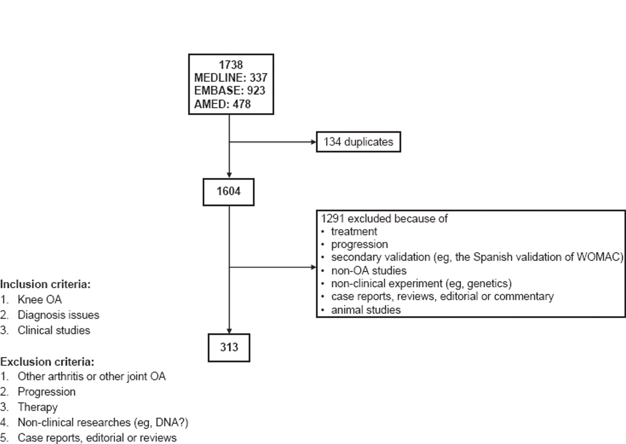
Study selection. OA, osteoarthritis; WOMAC, Western Ontario and McMaster Universities Osteoarthritis Index.
EULAR recommendations
Of 166 propositions suggested initially, 10 were agreed after four anonymous Delphi rounds. Recommendations covered the definition of knee OA and its risk factors, subsets, typical symptoms and signs, the use of imaging and laboratory tests and differential diagnosis (table 1). Evidence for validity (sensitivity, specificity, etc) and reliability of each diagnostic test/feature are summarised in table 2. Three symptoms (persistent knee pain, limited morning stiffness and reduced function) and three signs (crepitus, restricted movement and bony enlargement) appeared to be the most useful. Assuming a 12.5% background prevalence of knee OA in adults aged ≥45 years, the estimated probability of having radiographic knee OA increased with increasing number of positive features, to 99% when all six symptoms and signs were present (figure 2). Strength of recommendation was generated based on research evidence and clinical expertise with 95% CI (table 1). Details of each recommendation and supporting evidence are available online (supplementary file) in EULAR recommendations for the diagnosis of knee OA and supporting evidence.
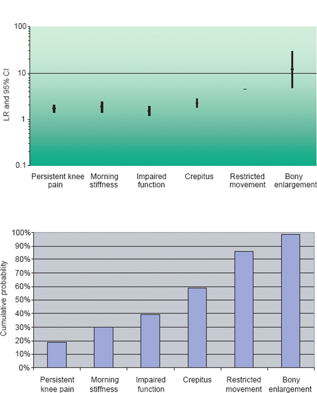
Likelihood ratio (LR) and probability of knee osteoarthritis (reference standard: radiographic KL ≥2). KL, Kellgren and Lawrence.
Propositions and strength of recommendation (SOR)—order according to topic (definition, subsets, symptoms, physical findings, images, laboratory tests, risk factors and differential diagnosis)
Validity and reliability of diagnostic tests in the diagnosis of knee osteoarthritis—pooled results
Performance of recommendations
The two populations selected had investigated plain radiographs and clinical features, permitting performance testing for some of the recommendations.
In the UK
We used cross-sectional data from the Knee Clinical Assessment Study (CAS(K)) conducted in North Staffordshire, UK. After excluding 16 people with a pre-existing diagnosis of inflammatory arthritis, 745 adults with knee pain aged ≥50 years (mean age 65 years, SD 8.6, range 50–93; 56% female; mean body mass index (BMI) 29.6 kg/m2, 41% obese) were available for analysis.22
Of 745, 570 (76%) and 292 (39%) subjects had radiographic OA according to two definitions based on standing posteroanterior, supine lateral and supine skyline views: osteophytosis (broadly equivalent to Kellgren and Lawrence score (KL) ≥1) and joint space narrowing (JSN) (broadly equivalent to KL ≥3). Compartmental distribution of knee OA differed according to the definition. With the first definition, the patellofemoral (PF) was the most commonly affected compartment (38%); with the second, the medial tibiofemoral (TF) was the most commonly affected (38%). The proportions with chondrocalcinosis in the same knee were 8% and 12%, respectively.
Age, gender, BMI, morning stiffness (<30 min), crepitus, reduced flexion, bony enlargement, fixed flexion deformity, palpable effusion and intercondylar/intermalleolar gap (a surrogate for varus/valgus malalignment) were entered in the logistic regression models and backward LR was used to select significant variables. Morning stiffness and intercondylar gap were excluded from the model because they were non-significant.
The probability of radiographic knee OA increased with an increasing number of positive tests (figure 3). The likelihood of having radiographic knee OA (KL≥1) was 88% for a person aged >60 years, who is overweight and has crepitus, restricted movement and bony enlargement. The likelihood was smaller when the diagnostic criterion was higher (eg, K L≥3) (figure 3).
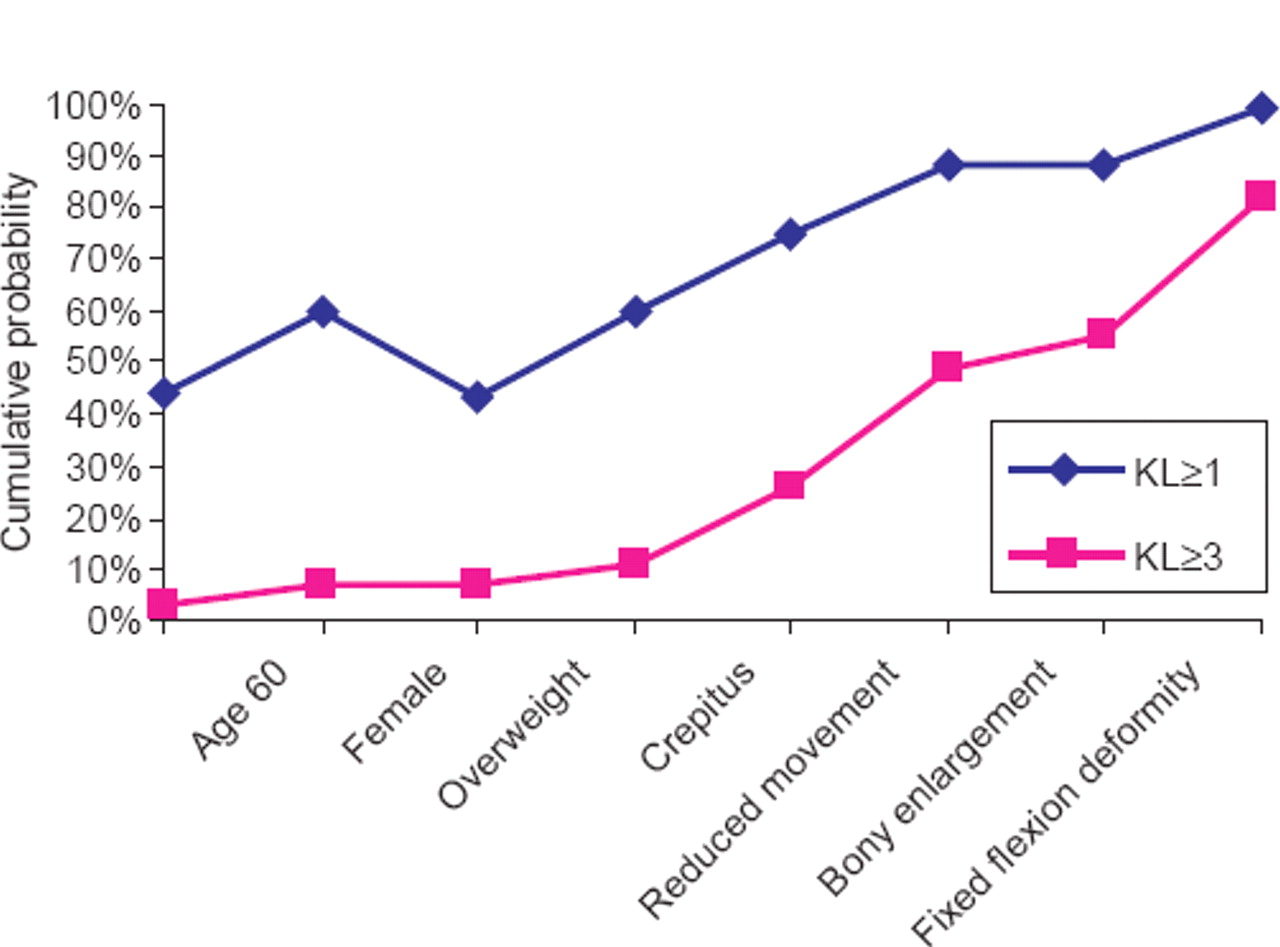
Clinical features and cumulative probability of radiographic knee osteoarthritis—evidence from the UK population. KL, Kellgren and Lawrence.
In the Netherlands
The Rotterdam study is a population-based, longitudinal cohort study for incidence and risk factors for chronic disabling conditions.52 Of 10 275 residents in one district of Rotterdam (Ommoord), 7983 agreed to participate (mean age 70.6, SD 9.8, range 55–106; 61.1% female, mean BMI 26.3, SD 3.7), 3456 with baseline knee anteroposterior (AP) x-rays formed the study population for this analysis.
Of 3456 subjects, 1624 (47%) and 129 (3.7%) were classified as having knee OA according to the cut-off points KL ≥1 and KL ≥3. Diagnostic variables examined included age, gender, BMI, knee pain in the past 5 years, morning stiffness, functional impairment, family history of OA, radiographic varus malalignment, hand OA (KL ≥2), hip OA (KL ≥2) and serum C-reactive protein (CRP) <5 mg/l. Of these, gender, morning stiffness, family history, hip OA and CRP were not significant so were excluded from the logistic regression models. Only four clinical features (age, BMI, knee pain and functional limitation) were available to test the performance of the clinical diagnosis. The probability of having any radiographic knee OA (KL ≥1) increased gradually with an increasing number of positive tests. It reached 52% when all these clinical features were positive—that is, aged >60 years of age, being overweight and having knee pain and impaired function.
Future research agenda
One hundred and thirteen initial propositions were submitted by the Task Force members. After three anonymous Delphi rounds, nine of these obtained over 50% votes and went forward as the proposed future research agenda:
(1) Development of internationally agreed criteria sets for diagnosis of knee OA for clinical practice, clinical trials and epidemiological studies.
(2) Development of a scoring system for accurate diagnosis of knee OA based on the sensitivity and specificity of risk factors and symptoms and signs.
(3) Delineation of the attributable risk factor profile, for both development and progression, for each suggested subset of knee OA.
(4) Development of diagnostic criteria for early symptomatic knee OA (eg, by prospective investigation of people with knee pain who fulfil criteria of knee OA several years later).
(5) Investigation of whether individual pain patterns (usage-related, episodic, night pain) have different utility as diagnostic markers of knee OA.
(6) Determination of clinical, diagnostic and prognostic relevance of MRI changes in knee OA.
(7) Determination of the utility of ultrasonography in the diagnosis and prognosis of knee OA.
(8) Assessment of the possible role of biomarkers (including genetic markers) in the early diagnosis, phenotypic characterisation and prediction of outcome of knee OA.
(9) Assessment of the accuracy of red flags in identifying serious pathology in patients presenting with knee symptoms.
Discussion
Knee OA can variably involve cartilage, bone, synovium and surrounding tissues of the three biomechanically discrete compartments and may associate with OA at other joints owing to shared genetic and constitutional risk exposures. Thus the clinical phenotype is very variable, requiring consideration of several characteristics for accurate diagnosis. Although the ACR criteria are a useful tool for classification of knee OA,11 they were developed using hospital-referred patients and a control group that comprised patients with other arthritis (over 50% had rheumatoid arthritis),11 thus making them most useful for differentiation of knee OA from inflammatory arthritis rather than for diagnosis of knee OA itself in a routine clinical setting. The focus of these current recommendations, however, was on the risk factors, symptoms, signs and tests that might contribute to a clinical diagnosis. Although there is no ‘gold standard’ for diagnosis of knee OA, an important conclusion was that in adults aged ≥45 years, an adequate history and examination alone may lead to a confident clinical diagnosis of knee OA. This is in contrast with the situation in some care settings, in which practitioners devote insufficient time to patient inquiry and physical examination and instead place undue emphasis on tests, especially radiographs.
The recommendations were developed systematically and combine both expert opinion (Delphi exercise) and research evidence (systematic review and meta-analysis).12 ,14 Evidence was derived from both community- and hospital-based studies to improve generalisability. The recommendations have been examined initially in datasets derived from two general populations in Europe.
According to the recommendations and the supporting evidence the diagnosis of knee OA can be made based on the background risk (the population prevalence of knee OA); the patient's risk factors for OA (eg, age, gender, BMI, occupation); their symptoms (persistent knee pain, brief morning stiffness and functional limitation) and an adequate physical examination (crepitus, restricted movement and bony enlargement). Plain radiographs are the main test to consider, but are an adjunct, rather than a central feature, for the purposes of diagnosis (figure 4). The more positive results a patient presents, the more likely the diagnosis of OA. Knowledge of the background risk (ie, the local source population prevalence of knee OA) is crucial for estimating the likelihood of knee OA. The higher the risk in the source population, the more possible it is to diagnose knee OA based on clinical features.

Major components in the diagnosis of knee osteoarthritis (OA). BMI, body mass index.
There are limitations to these recommendations. First, the evidence to support these recommendations was derived largely from literature based on different studies. The LRs (table 2) are unadjusted and the subsequent probabilities are for reference only. The application of these recommendations should be based on the individual patient characteristics and the knee OA risk in the source population. Second, the LRs pooled from the literature may be affected by many factors, such as the number of studies involved, the populations selected (hospital or community), the ‘gold standard’ used and the cut-off values selected. For example, the LR for bony enlargement (11.81, 95% CI 4.94 to 28.22) was mainly based on a hospital-based case–control study where the ‘gold standard’ was clinical diagnosis of knee OA and the controls predominantly were patients with rheumatoid arthritis.11 The validity and reliability of this LR is questionable, compared with those LRs derived from multiple studies including both hospital and community data, such as for persistent knee pain28,–,30 and crepitus.27 ,32 ,34 Therefore caution must be exercised when interpreting results obtained using this feature. Third, there is no universally applicable reference standard for knee OA, so the recommendations were mainly based on radiographic evidence when clinical features were examined, or on clinical, MRI or arthroscopic evidence when radiographic features were examined. Whether this is an appropriate approach is open to debate. Finally, all propositions relate to people over age 40, which is the target age for common OA. Whether recommendations would differ for less typical patients under this age was not examined.
In conclusion, 10 key recommendations for the diagnosis of knee OA have been produced based both on expert consensus and a systematic literature review. A confident diagnosis may be made according to three symptoms (knee pain, short-lived morning stiffness and functional limitation) and determination of three signs on examination (crepitus, restricted movement and bony enlargement) without a requirement for imaging. This may be especially useful for primary care. Nevertheless, plain radiography and occasionally other investigations may be considered for the diagnosis of atypical cases when additional pathology is suspected. These recommendations were examined in two test populations and the level of evidence and summary strength of recommendations were provided to guide their use.
Acknowledgments
We thank the European League Against Rheumatism for financial support, Helen Richardson for co-ordination and Joanna Ramowski for assistance with the literature search and paper collection.
References
Supplementary materials
Web Only Data ard.2009.113100
Files in this Data Supplement:
Footnotes
-
Funding Financial support was received from the European League Against Rheumatism.
-
Competing interests None.
-
Provenance and peer review Not commissioned; externally peer reviewed.