Article Text

Abstract
Background Previous studies on the prognostic value of clinical and MRI parameters for the time to return to play (TTRTP) in acute hamstring injuries showed only limited to moderate evidence for the various investigated parameters. Some studies had multiple methodological limitations, including retrospective designs and the use of univariate analysis only. The aim of this study was to assess the prognostic value of clinical and MRI parameters for TTRTP using multivariate analysis.
Methods 28 clinical and MRI parameters were prospectively investigated for an association with TTRTP in 80 non-professional athletes with MRI positive hamstring injuries undergoing a standardised rehabilitation programme. The association between possible prognostic parameters and TTRTP was assessed with a multivariate linear regression model. Parameters that had a p value <0.2 on univariate testing were included in this model.
Results 74 athletes were available for analysis. A total of nine variables met the criteria for the multivariate analysis: intensity of sports, level of sports, self-predicted TTRTP by the athlete, length of discomfort on palpation, deficit in passive straight leg raise, pain score on isometric knee flexion, isometric knee flexion strength deficit and distance of the proximal pole of the MRI hyperintensity to the tuber ischiadicum. Of these, only self-predicted TTRTP by the athlete and a passive straight leg raise deficit remained significantly associated with TTRTP after stepwise logistic regression.
Conclusions The clinical parameters self-predicted TTRTP and passive straight leg raise deficit are independently associated with the TTRTP. MRI parameters in grade 1 and 2 hamstring injuries, as described in the literature, are not associated with TTRTP. For clinical practice, prognosis of the TTRTP in these injuries should better be based on clinical parameters.
- Hamstring
- MRI
- Soft tissue
- Muscle damage/injuries
Statistics from Altmetric.com
Introduction
After acute hamstring injury, the primary question of the athlete, medical and coaching staff is how long it will take to return to play. The large variation of 1 day1 to 104 weeks2 in time needed to return to play (TTRTP) makes estimating the prognosis a challenge. Few studies have evaluated the prognostic value of findings on clinical assessment for the TTRTP, showing limited evidence that a visual analogue pain score of the injury,3 time taken to walk pain free4 and stretching mechanism of injury are associated with the TTRTP.
A substantial number of studies have identified possible prognostic MRI parameters.3 ,5–15 There is only limited to moderate evidence for an association of a hyperintensive signal on T2-weighted images, involvement of the proximal or central tendon, injury not affecting the musculotendinous junction and a total rupture with a longer TTRTP. 3 ,5–15
Methodological limitations of some of these studies are the relative low number of participants, retrospective study designs, lack of blinding and the use of simplistic univariate statistical analysis. As none of the studies used multivariate analysis in which both clinical and MRI findings were analysed, it remains unknown to what extent MRI findings are independently associated with the TTRTP and complementary to clinical predictors. Additionally, in none of the studies were athletes or decisionmakers for return to play blinded for the clinical or MRI assessment, introducing a substantial risk of bias.
Therefore, the aim of this study was to assess the prognostic value of clinical and MRI parameters for the TTRTP after acute hamstring injury. For this objective, a prospective design, multivariate analysis and blinding of athletes and decisionmakers for return to play were ensured.
Methods
Participants
The athletes in this study took part in a previously published multicentre randomised controlled trial on the effect of platelet rich plasma in hamstring injuries (Dutch trial register number 2771). This trial started in and was conducted between February 2011 and May 2013 at the sports medicine departments of a general district hospital, a university hospital and at the FIFA medical centre of excellence of the national football association in the Netherlands. In this study, athletes were randomised into either an intervention group or a control group. The intervention group received two injections of 3 mL platelet-rich plasma (Autologous Conditioned Plasma, Biocore, Arthrex Inc, Karlsfeld, Germany) and the control group received two injections of 3 mL saline at the site of the injury. The first injection was performed within 5 days of the injury and the second injection 5–7 days later. Injections were performed using a sterile ultrasound-guided technique into the region of maximal muscle injury determined by MRI. All athletes completed a standardised physiotherapy programme, including range of motion exercises, progressive strength exercises, core stability training and agility exercises.16 ,17 The exercises were all supervised by a specially instructed sports physiotherapist. There were no differences between the intervention and the control group on the primary outcome measure of the TTRTP.
All athletes provided written informed consent prior to the start of the study. Approval was obtained from the Regional Ethical Committee of South West Holland.
Eligibility criteria
Athletes were included if they met the following criteria: age of 18–50 years; a clinical diagnosis of an acute hamstring injury defined as a history of acute posterior thigh pain within the past 5 days, localised discomfort on palpation, localised pain on passive stretching of the hamstrings and increased pain on isometric contraction of the hamstring; and a visible hamstring lesion on MRI (within 5 days of injury), defined as an increased signal on fluid sensitive sequences.
Athletes were excluded if they were not capable of performing an active exercise programme; if they had already received an injection for the injury; if they had no intention to return to full sports activity; if they did not want to receive one of the two therapies in the trial; if the cause of the injury was an extrinsic trauma (contusion injury); if they had chronic hamstring symptoms, defined as recurrent tenderness of the hamstring muscles in the previous 2 months; if they had chronic low back pain; if they had a contraindication for MRI; or if there was a total rupture and/or avulsion seen on MRI.
Baseline assessment
All baseline assessments were performed on the same day within 5 days of the occurrence of the injury and before any injections were given.
Questionnaire
Patient characteristics, level and intensity of sports participation, information on history of previous hamstring injuries, history of anterior cruciate ligament surgery using a hamstring graft, the injury mechanism, the ability to walk pain free within 1 day and the self-predicted days to RTP indicated by the patient were obtained using a structured questionnaire.
Clinical examination
Manual muscle palpation
With the patient in a prone position the complete posterior thigh was carefully palpated from the hamstring origin at the ischial tuberosity to the insertions medial at the pes anserinus and lateral at the head of the fibula. The total longitudinal length of the discomfort area, the distance between the proximal border of the discomfort area on palpation of the hamstrings and the ischial tuberosity, and the distance between the point of maximal discomfort on palpation and the ischial tuberosity were recorded.
Hamstring flexibility testing
Hamstring flexibility was assessed with the active knee extension test18 ,19 and the passive straight leg raise test.20 Athletes were tested in a supine position with an inclinometer placed on the anterior border of the tibia. For the active knee extension test athletes were asked to position the hip of the tested leg in 90° flexion and instructed to extend the knee until maximal tolerable stretch, with the contralateral leg remaining flat on the examination table. At the endpoint of maximal tolerable stretch, the absolute knee angle was measured. For the passive knee extension test athletes were instructed to completely relax the leg, while the researcher lifted the leg with the knee in full extension until maximal tolerable stretch, with the contralateral leg remaining flat on the examination table. At the endpoint of maximal tolerable stretch, the angle between the leg and the horizontal was measured. For both tests the absolute flexibility deficit was calculated by subtracting the recorded angle of the injured leg from the uninjured leg. Additionally, athletes were asked whether they experienced normal stretch or localised pain during the tests.
Isometric knee flexion force
Isometric knee flexion force was measured using handheld dynamometry.21 Athletes were tested in a prone position with the knee in 15° of knee flexion. The researcher placed the dynamometer at the heel of the participant and applied force to the heel, gradually increasing in 3–5 s. Athletes were instructed to resist the force applied by the researcher (brake test). At the point that the participant could not resist the force any more the test was terminated and the dynamometer was read out. Each leg was tested three times. For each angle the highest force value was recorded. The relative strength deficit was calculated by dividing the recorded maximal force value of the injured leg by maximal force value of the uninjured leg. Additionally, athletes were asked whether they experienced localised pain during the test.
MRI assessment
The used protocol was a modified version of the protocol described by Askling et al.5 To locate the area of the injury, the entire hamstring of the injured limb was visualised by obtaining coronal and sagittal short τ inversion recovery (STIR) images from the ischial origin of the hamstring muscles to the insertion on the fibula and the tibia (repetition time/echo time (TR/TE) of 3500/31 ms, field of view (FOV) of 300 mm and a 256×320 matrix). The uninjured leg was not depicted. Subsequently, transversal STIR (repetition time/echo time (TR/TE) of 3500/31 ms, FOV of 300 mm and a 205×256 matrix), T1-weighted (TR/TE of 500/12 ms, FOV of 300 mm and a 355×448 matrix) and T2-weighted (TR/TE of 4080/128 ms, FOV of 300 mm and a 355×448 matrix) images were obtained from the injured area. The thickness of the slices for all sequences was 5 mm. MRIs were obtained with a 1.5-T magnet system (Magnetom Essenza, Siemens) with the use of a body matrix coil.
Each MRI was assessed by one radiologist, specialised in musculoskeletal radiology, who was blinded for all information except that there was a clinical diagnosis of a hamstring injury. For assessment of the MRIs we used standardised scoring forms based on the literature.5 ,9 ,11 ,15 ,22 We recorded the involved muscle(s) and performed grading of the injury using the three-graded classification of Hancock et al22: grade (1): increased signal intensity on fluid sensitive sequences without evidence of a macroscopic tear, grade (2): increased signal intensity on fluid sensitive sequences with a partial tear, and grade (3): total muscle or tendon rupture. When no abnormalities were found, we regarded this as a grade 0 injury. We measured the increased T2 signal intensity for the affected hamstring muscle in craniocaudal, transverse and anteroposterior dimensions on the fluid sensitive sequences (STIR). Increased signal intensity was defined as an abnormal intramuscular increased signal compared to the unaffected surrounding muscle tissue. We recorded the longitudinal length (craniocaudal) and calculated the involved cross-sectional area as a percentage of the total muscle cross-sectional area in the transversal plane and the total volume using the formula of a prolate ellipsoid (4/3π×length×width×depth). We measured the distance of the most cranial pole of the intramuscular increased signal intensity to the distal tip of the ischial tuberosity and recorded whether there was extramuscular fluid present. Good to excellent interobserver and intraobserver reliability was found for the used MRI parameters in a previous study.23
Outcome measure
The outcome was the time to return to play (TTRTP), defined as the number of days between injury and return to unrestricted sports activity in training and/or match play.24 On a daily basis the athletes performed in a progressive phased, criteria-based rehabilitation programme, which was based on the best available evidence.16 ,17 Patients were instructed to contact the coordinating researcher at the moment of return to unrestricted sports activity. The definite clearance for RTP was given by the supervising physiotherapist once the patient completed the rehabilitation programme, including unrestricted functional sport specific testing. The athletes and the supervising physiotherapists were blinded to the clinical and MRI parameters assessed at baseline. We contacted athletes that did not return to play yet at 1, 3, 4, 8, 10, 16 and 26 weeks after inclusion to assess TTRTP. Athletes who sustained another non-hamstring injury before RTP were excluded from the analysis.
Statistical analysis
We performed statistical analyses with SPSS software (V.20.0; SPSS, Chicago, Illinois, USA). We analysed baseline patient characteristics using descriptive statistics. If the data were normally distributed, continuous variables were presented as a mean with a SD, otherwise a median and IQR were used.
We analysed the association between the possible predictive variables measured at baseline and the TTRTP with a linear regression model. Variables that had a p value <0.2 on univariate testing were included in a multivariate backward linear regression model. We used a probability of F for removal of 0.10. We calculated adjusted regression coefficients (β-coefficients) and 95% CIs for the included predictive variables. Finally, the total variance of these predictive variables for TTRTP explained by the model was calculated.
Results
Study patients and follow-up
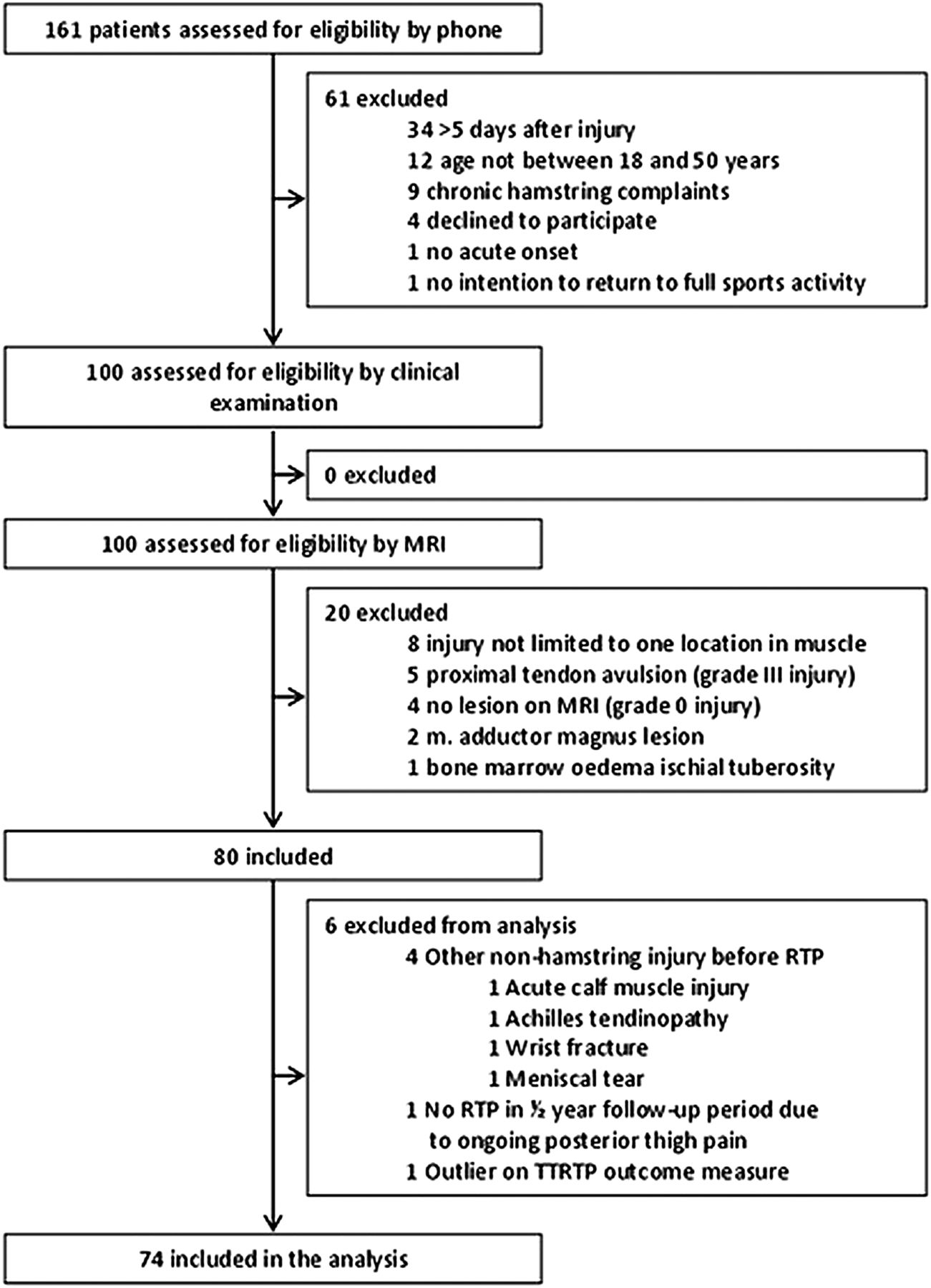
Between February 2011 and November 2012, 80 patients were included. Five patients did not achieve RTP within the study period and were excluded from the analysis: four patients sustained another non-hamstring injury before RTP, one patient did not manage to RTP because of ongoing posterior thigh symptoms. There was one participant with a time to RTP of 149 days who was considered an outlier and therefore excluded from the analysis (figure 1). Of the 74 patients included in the analysis the sports played were football (n=55, 74%), field hockey (n=12, 16%), track and field athletics (n=4, 5%), fitness (n=1, 1%), American football (n=1, 1%) and tennis (n=1, 1%). The majority of the athletes played on a non-professional, competitive level (74%), the other athletes competed recreationally. The median time between injury and baseline assessment was 3 days (IQR, 2–4). Other baseline characteristics are presented in table 1. The mean time to RTP was 44 days (±18).
Baseline assessment and their association with time to return to play in univariate analysis

Patient flow diagram.
Association of clinical and MRI assessment with TTRTP
The association of the baseline assessment with the TTRTP analysed with univariate linear regression model is presented in table 1. There were nine variables with a p value <0.2 that were included in the multivariate analysis: intensity of sports, level of sports, self-predicted TTRTP by the athlete, length of discomfort on palpation, passive straight leg raise deficit, isometric knee flexion strength testing pain score, isometric knee flexion strength testing force deficit and distance of proximal pole of the hyperintensity seen on MRI.
After backward regression, three variables were included in the model, of which two were independently statistically significantly associated with the TTRTP (see table 2): the deficit in passive straight leg raise in degrees (β-coefficient 0.70; 95% CI 0.13 to 1.27; p=0.017) and the self-predicted TTRTP indicated by the patient in days (β-coefficient 0.36; 95% CI 0.01 to 0.71; p=0.045). The variance in TTRTP explained by the model was 20% (R2=0.20).
Multivariate analysis
Discussion
This prospective study on acute hamstring injury revealed after multivariate analysis that only athletes’ self-predicted TTRTP and passive straight leg raise deficit were independently associated with TTRTP. None of the MRI parameters were independently associated with the TTRTP. Our findings reflect the value of clinical parameters.
Clinical parameters
Most of the previous studies on the value of clinical predictive parameters used univariate analysis. In absence of multivariate analysis it remains unknown to what extent the predictive parameters are independently associated with the time to RTP, as the majority can be expected to be mutually correlated. To assess the different clinical parameters independently, we used multivariate analysis.
Owing to the limited number of studies examining clinical parameters for their prognostic value, the possibility to compare our results with findings in the literature is limited. The value of self-predicted TTRTP was assessed in one study, although unpublished.25 Eighteen athletes (sprinters) self-estimated the time to be back at the preinjury level. The self-predicted time to return at preinjury level was 4 weeks (median, range 2–12), while the actual time to return was significantly longer (median 16 weeks, range 6–50). However, no measure of association between self-predicted time to be back at preinjury level and TTRTP was reported. Our study found a significant association between self-predicted TTRTP and reported TTRTP. A possible explanation might be that over 60% of the athletes had a previous hamstring injury. This previous experience might be used by the athlete as a reference standard, possibly leading to bias.
Additionally, a previous study showed that the predicted TTRTP by a sports physician based on clinical examination was as good as predicting TTRTP with MRI, providing more leverage for clinically assessing TTRTP.10 Contrary to the two previous studies, we found that passive straight leg raise deficit was significantly associated with TTRTP.4 ,20 Warren et al, not finding such an association, investigated 59 Australian Football players with an acute hamstring injury and used stepwise logistic regression. Possibly, the different findings of Warren et al are due to methodological differences. Warren et al4 assessed a deficit in passive straight leg raise dichotomously (≤10° and >10°), while we scored continuously. Additionally, our study included athletes with an MRI positive, while Warren et al included clinically positive injuries. Askling et al20 reported no statistical significant association between a deficit in passive straight leg raise and TTRTP in 18 elite sprinters and 15 professional dancers. The absence of association might potentially be caused by the relative low sample size.
Several other clinical parameters were reported in the literature to be associated with TTRTP. The significant associated parameters reported were: time to walk pain free,4 active knee extension deficit >10°, discomfort on hamstring palpation localised more cranial to the tuber ischii,5 ,7 stretching type hamstring injury7 ,20 and maximum pain experienced with the injury.3 However, in these studies, the sample size was usually small, outcome assessors were not blinded for the studied prognostic parameter and/or no multivariate analysis was used.3 ,5 ,7 ,19 ,20 The reported association was not confirmed in our cohort.
Overall, the estimated TTRTP and deficit in passive straight leg raise explained only 20% of the total explained variance. To describe the clinical relevance of our findings an example for clinical practise is provided below.
The mean TTRTP for the group was 44±18 days, indicating that approximately 95% of the athletes returned to play between a range of 8 and 80 days (mean ±2 times the SD). With the self-predicted and passive straight leg raise deficit we could only narrow the range down slightly. For an athlete, with a self-estimated TTRTP of 42 days and a passive straight leg raise deficit of 10°, the 95% CI for the estimated TTRTP by the model is 16–83 days, instead of 8–80 days. This wide CI implies that future studies are needed to reveal additional prognostic parameters to increase the percentage of the explained variance.
MRI parameters
In this study, patients and decisionmakers for RTP were blinded for the MRI results, thus keeping the risk of bias low. In other studies on the prognostic value of MRI parameters in acute hamstring injuries, this blinding was not ensured or not described. Multiple studies have investigated the association between one or more MRI parameters in acute hamstring injuries and TTRTP,3 ,5–9 11–15 with correlation coefficients ranging from 0.39 to 0.74 to assess the extent of the injury. In these studies, blinding of the patients and decisionmakers for RTP was not ensured or not described.
None of these studies used a multivariate analysis and therefore it remains unknown to what extent the MRI parameters have any prognostic value additional to the parameters obtained by clinical evaluation. The findings of our study suggest that the prognostic capacity of an MRI scan for acute hamstrings injuries might not be as strong as previously stated in the literature. In the present study, none of the MRI parameters were significantly associated with TTRTP after multivariate analysis.
We have to emphasise that in our study no hamstrings were included that showed no abnormalities (grade 0) on MRI. Also, we excluded total hamstring ruptures (grade 3). Therefore, only grade 1 and 2 lesions were included.22 Ekstrand et al11 found that grade 0 lesions had a shorter TTRTP than grade 1 and 2 lesions. They also found that grade 1 and 2 lesions were the most common grades of injury, (respectively, 57% and 27% of the total hamstring injuries). In addition, they found that grade 3 lesions displayed the longest TTRTP. Owing to the nature of our inclusion criteria (MRI positive hamstring injuries (grade 1 and 2) and exclusion of total ruptures (grade 3)), no comparison with the grade 0 and grade 3 injuries from the Ekstrand et al11 study was possible.
When comparing our results with findings in the literature, Hallen and Ekstrand,26 in the large UEFA and Champions League study, did find a difference in TTRTP between grade 1 and grade 2 injuries on MRI (median 15 days for grade 1 injuries (IQR 14 days) and median 21 days for grade 2 injuries (IQR 19 days); p<0.0001). This difference can possibly be explained by difference in the study population (professional vs non-professional), the number of participants and the statistical analysis used in the studies.
Unfortunately, no comparison between the prognostic value of the involvement of the free proximal tendon was possible. In two studies that investigated the prognostic value of the involvement of the proximal free tendon, comparison with the uninjured leg was used.5 ,6 Our limitation is that we only depicted the injured leg, which excluded direct comparison. For the future, the association of involvement of the free proximal tendon and TTRTP should be investigated more extensively. Possibly, new MRI techniques, such as 3 Tesla scans might enhance the prognostic value of MRI scans.
Strength of the study
This study has several strengths. First, the size of the athletic population was quite large and the design of the study was prospective. Second, multivariate linear regression was used to examine the possible prognostic parameters for their independent association. Furthermore, treatment of the athletes was not influenced by the baseline characteristics, since treatment was allocated randomly. In addition, the decisionmaker for TTRTP was blinded to the baseline characteristics, including MRI.
Limitations
Although 74 athletes were included in this study, there could be a lack of power to detect weak associations with TTRTP. Owing to the fact that no professional athletes were included in the study, caution has to be taken in generalising the results of this study to a professional athletic population. As no grade 0 and grade 3 injuries were included in this study, no comparison with other studies that looked at the association of the different grades of injury of the hamstring, and our study, was possible.
Conclusion
The clinical parameters of self-predicted TTRTP and passive straight leg raise deficit are independently associated with the TTRTP. MRI parameters in grade 1 and 2 hamstring injuries, as described in the literature, are not associated with TTRTP. For clinical practice, prognosis of the TTRTP in these injuries should better be based on clinical parameters.
What are the new findings?
-
Clinical parameters self-predicted time to return to play (TTRTP) and passive straight leg raise deficit in grade 1 and 2 acute hamstring injuries are associated with TTRTP in non-professional athletes, while MRI parameters are not.
How might it impact on clinical practise in the near future?
-
It is advised to evaluate a passive straight leg raise test and self-predicted TTRTP to guide the prognosis. In non-professional athletes, performing an MRI to estimate the TTRTP is not routinely advised.
References
Footnotes
MM and GR contributed equally to this manuscript.
-
Contributors MHM and GR designed the study, monitored data collection, analysed and interpreted the data and revised the paper. JLT, AW, MHM and GJG interpreted the data and revised the paper.
-
Funding The randomised controlled trial was supported by the Royal Dutch Football Association and Arthrex Medizinische Instrumente GmbH.
-
Competing interests None.
-
Patient consent Obtained.
-
Ethics approval Approval was obtained from the Regional Ethical Committee of South West Holland.
-
Provenance and peer review Not commissioned; externally peer reviewed.