Article Text

Abstract
Background Proximal muscle rehabilitation is commonly prescribed to address muscle strength and function deficits in individuals with patellofemoral pain (PFP). This review (1) evaluates the efficacy of proximal musculature rehabilitation for patients with PFP; (2) compares the efficacy of various rehabilitation protocols; and (3) identifies potential biomechanical mechanisms of effect in order to optimise outcomes from proximal rehabilitation in this problematic patient group.
Methods Web of Knowledge, CINAHL, EMBASE and Medline databases were searched in December 2014 for randomised clinical trials and cohort studies evaluating proximal rehabilitation for PFP. Quality assessment was performed by two independent reviewers. Effect size calculations using standard mean differences and 95% CIs were calculated for each comparison.
Results 14 studies were identified, seven of high quality. Strong evidence indicated proximal combined with quadriceps rehabilitation decreased pain and improved function in the short term, with moderate evidence for medium-term outcomes. Moderate evidence indicated that proximal when compared with quadriceps rehabilitation decreased pain in the short-term and medium-term, and improved function in the medium term. Limited evidence indicated proximal combined with quadriceps rehabilitation decreased pain more than quadriceps rehabilitation in the long term. Very limited short-term mechanistic evidence indicated proximal rehabilitation compared with no intervention decreased pain, improved function, increased isometric hip strength and decreased knee valgum variability while running.
Conclusions A robust body of work shows proximal rehabilitation for PFP should be included in conservative management. Importantly, greater pain reduction and improved function at 1 year highlight the long-term value of proximal combined with quadriceps rehabilitation for PFP.
- Exercises
- Knee
- Rehabilitation
Statistics from Altmetric.com
Introduction
Patellofemoral pain (PFP) is one of the most common presentations at both primary care and sports injury clinics.1 ,2 Prevalence rates in groups of active individuals, including military recruits and novice runners, are reported to be between 3% and 20%.3–5 PFP has been linked to reduced contact area and increased stress in the lateral patellofemoral joint (PFJ)6 ,7 as a result of patellar maltracking, including greater lateral patellar translation,8–10 tilt8 and spin.9 The cause of maltracking in PFP is thought to be multifactorial with local,11 distal12 and proximal13 factors proposed to contribute to it, with good evidence that long axis femoral rotation in relation to the patella is a key contributor to maltracking and a valid rehabilitation target.14
Consistent with the multifactorial nature of PFP, management of PFP has traditionally focused on a variety of interventions, including rest, analgesia, general quadriceps and vastus medialis oblique rehabilitation exercises, proximal rehabilitation exercises, patellar taping, foot orthoses and gait retraining.15 Each of these interventions has a varying level of efficacy, with multimodal interventions appearing to be the most effective.16 ,17 Growing evidence for impaired proximal muscle strength18–20 and function,21 combined with links between hip mechanics and increased risk of PFP,5 ,22 has resulted in promotion of rehabilitation aimed at addressing impairments in proximal musculature.14 Our recent mixed methods study of international experts’ clinical reasoning when managing PFP supported this recommendation, but a lack of supporting level-one evidence was also identified.17 The effectiveness of proximal rehabilitation protocols have been evaluated in high quality recent research,23–26 and commonly consist of open and closed kinetic chain exercises which reflect clinical practice.27
A recent low quality (LQ) systematic review concerning proximal rehabilitation for PFP28 concluded that hip interventions were effective in improving pain and function in individuals with PFP. However, the search for available evidence was limited to a 2-year period (January 2011–January 2013), with no attempt at data pooling nor mechanistic exploration and there is, therefore, a need for a more detailed and inclusive review in order to optimally guide practice.
Our systematic review and meta-analysis aims to (1) evaluate the effects of proximal muscle rehabilitation for patients with PFP, (2) compare the effects of various rehabilitation protocols, and (3) evaluate potential mechanism of action in order to optimally guide clinical practice in rehabilitating patients. Further, we aimed to promote clarity in rehabilitation programme design and reporting, with particular respect to the term ‘strengthening’.
Methods
The PRISMA statement was consulted prior to the start of this review and the checklist completed.29
Inclusion and exclusion criteria
Randomised clinical trials (RCTs) and cohort studies evaluating proximal muscle rehabilitation programmes were considered for inclusion. A proximal muscle rehabilitation protocol was defined as progressive exercise directed at the hip or lumbopelvic musculature or both. Studies of multimodal interventions were included where the effects of adding proximal rehabilitation could be clearly determined. Case reports and non-English studies were excluded. The inclusion criteria required participants to be described as having PFP, anterior knee pain or chondromalacia patella in the absence of other knee pathologies, including patellar tendinopathy, Osgood-Schlatters disease and Sinding-Larsen-Johanssons syndrome. Studies evaluating all age ranges were considered for inclusion, as well as studies involving both single sex and mixed sex sample groups.
Search strategy
Web of Knowledge, CINAHL, EMBASE and Medline (via OVID) databases were searched from inception to December 2014, using the search strategy outlined in box 1. Reference lists of included publications were screened and citation tracking was completed in Google Scholar.
Search strategy
Patellofemoral Pain OR Anterior Knee Pain OR Patellofemoral Syndrome OR retropatellar pain OR peripatellar pain OR patellofemoral joint pain OR parapatellar pain OR PFP OR chondromalacia patellae
AND
Proximal OR gluteal
AND
Strength*
AND
Training OR program OR exercise OR rehab*
*, a truncation indicator for searching.
Review process
Titles and abstracts identified using the search strategy were downloaded into EndNote X7.1 (Thomson Reuters, California, USA). Duplicates were deleted before all abstracts were screened for inclusion by two independent reviewers (SL and OS). A third reviewer (CB) was available to settle any disputes if necessary. Full texts were obtained where necessary.
Quality assessment
Study methodological quality was assessed with the PEDro scale30 and a PFP inclusion/exclusion criteria checklist31 by two independent reviewers (OS and SL). Discrepancies were resolved by consensus, with a third reviewer (CB) available, if needed. Based on the PEDro scores,30 and guidance by Moher et al,32 studies scoring >6 were considered high quality (HQ) and ≤6 as LQ. The PFP diagnosis checklist31 is a seven-item scale that identifies key inclusion and exclusion criteria for the diagnosis of PFP. Higher scores indicate a greater number of key criteria having been reported.
Study analysis
Sample sizes, participant demographics, interventions, variables evaluated and follow-up times were extracted from each study. Further analysis of intervention programme design was completed to determine the type of ‘rehabilitation’ that was prescribed (table 2) allowing for direct comparison between reported training methodology and accepted principles of ‘neuromuscular activation’ (exercise performed at <30% 1 repetition maximum, for >20 repetitions), ‘strength’ (exercise performed ≥70% 1 repetition maximum), ‘strength-endurance’ (exercise performed at 30–70% 1 repetition maximum) and ‘power’ (exercise performed at either 85–100% 1 repetition maximum or 0–60% 1 repetition maximum at an explosive velocity).33 This was in response to concerns raised at the recent PFP research retreat in Vancouver15 that the word strengthening or strength training is used synonymously for all types of rehabilitation exercise consequently limiting the identification of exercise prescription specificity. It was considered that evaluating methods of exercise prescription (eg, focus on strength, endurance, etc.) and summarising the range of specific exercise descriptors (% repetition maximum, repetitions, time-under-tension) could maximise the clinical utility of this review and facilitate translation to clinical practice.
Means and SDs for all baseline and follow-up data were extracted and entered into Cochrane Review Manager (V.5.2) to allow calculation of standard mean differences (SMDs). Meta-analysis was completed where studies evaluated similar interventions using comparable outcome measures (eg, VAS, Visual Analogue Scale and NPRS, Numeric Pain Rating Scale). Where multiple measures were used, a consistent measure between studies was used for pooling (eg, stair ascent). Pooling of data across time points was performed for studies that evaluated outcomes in the ‘short term’ (<3 months), ‘medium term’ (3–12 months), and ‘longer term’ (≥12 months). All outcome measure scores were converted so that favourable outcomes (reduced pain, improved function, improved strength, etc) were entered as positive values into Cochrane Review Manager Software, to facilitate consistent visual representation of SMDs and pooled findings along with 95% CIs. For studies without comparative groups, the results were extracted and reported in the results section, but no meta-analysis performed. Following methodology proposed by Hume et al,34 individual and pooled SMDs were categorised as small (≤0.59), medium (0.60–1.19), or large (≥1.20). These criteria were chosen to increase stringency compared to traditional criteria.35 Statistical heterogeneity of pooled data was established using the X2 and I2 statistics (with heterogeneity being defined as p<0.05). Levels of evidence were guided by recommendations proposed by Van Tulder et al:36
Strong evidence=based on results derived from multiple studies, including a minimum of two HQ studies, which are statistically homogenous (I2<50%).
Moderate evidence=based on results derived from multiple studies, including at least one HQ study, which are statistically heterogeneous (I2>50%), or from multiple LQ studies which are statistically homogenous (I2<50%).
Limited evidence=based on results derived from multiple LQ studies which are statistically heterogeneous (I2>50%), or from one HQ study.
Very limited evidence=based on results derived from one LQ study.
Conflicting evidence=based on insignificant pooled results derived from multiple studies regardless of quality, which are statistically heterogeneous (I2>50%).
Results
The results of the database search are shown in figure 1. Fourteen studies were identified for the final review. Eleven of these studies were randomised or comparative control trials,23 ,24 ,26 ,37–44 and three were cohort studies.25 ,45 ,46 Study details, including sample sizes and participant demographics, are shown in online supplementary file 1. Intervention and control/comparison group protocols, outcome measures and follow-up duration findings are presented in online supplementary files 2–5.

Flow chart of included studies. PFP, patellofemoral pain.
Quality assessment
Results from the PFP diagnostic checklist and the PEDro scale are shown in online supplementary file 6 and table 1, respectively. All 12 studies scored five or greater out of seven on the PFP diagnostic checklist, demonstrating a good level of consistency between studies for diagnostic inclusion/exclusion criteria. Scores ranged between 3 and 10 for the PEDro scale. Of the 14 studies, 7 were classed as HQ,23 ,38–40 ,42–44 and 7 were classed as LQ.24–26 ,37 ,41 ,45 ,46
PEDro scale
Exercise prescription and mechanobiological analysis
Results of exercise prescription and mechanobiological analysis are shown in tables 2 and 3, respectively. Three of the 14 included studies23 ,38 ,44 were considered to have evaluated the same exercise approach as stated in their title and methodology. Commonly (10 of 14 studies), studies reported evaluation of a strength protocol, despite exercise programmes being considered to be of an intensity to evoke strength-endurance24 ,37 ,39 ,42 ,45 ,46 or neuromuscular25 ,26 ,40 ,43 activation changes. In one study, the description of exercise prescribed was unclear and could not be interpreted.41 Analysis of mechanobiological descriptors of exercise prescription from within the 14 included studies highlighted the absence of all ‘classical descriptors’ (eg, load magnitude, % of maximum) and rest period between sets (s/min)) in all but one study39 (table 3). The seven new descriptors proposed by Toigo and Boutellier47 were absent, in their entirety, in the methodology of all included studies. Inclusion of the 13 descriptors in future studies is reported to be imperative for the delivery of effective and tailored exercise prescription.47
Analysis of programme design and aims
Analysis of specific descriptors of exercise prescription
Effects of proximal rehabilitation
Proximal rehabilitation—compared with—control
Pain and function
One LQ study26 compared proximal rehabilitation in PFP patients to a control group receiving only Omega-3 and calcium supplementation in the short term (figure 2). Very limited evidence (1 LQ study26) with large effect indicated proximal rehabilitation, using exclusively open kinetic chain (OKC) exercises with progressively higher resistant elastic band (see online supplementary file 2 for further programme details), reduces pain (VAS) (SMD, 95% CI 2.80,1.71 to 3.88), and improves function (Western Ontario and McMaster osteoarthritis index (WOMAC)) (SMD, 95% CI 2.88, 1.78 to 3.98) in the short term.

Proximal rehabilitation compared with control group for pain and function (WOMAC, Western Ontario and McMaster osteoarthritis index; ‘short term’, <3 months; IV, inverse variance; Std., standard mean difference).
Proximal rehabilitation—compared with—quadriceps rehabilitation
Pain
Three HQ42–44 and one LQ study24 compared proximal rehabilitation to quadriceps rehabilitation in the short and medium term (see online supplementary file 3 for further programme details; figure 3). Moderate evidence (3 HQ42–44 and 1 LQ24 study) of a small effect indicated greater pain reduction following a proximal rehabilitation programme compared with a quadriceps rehabilitation protocol in the short term (I2=81%, p=0.001; SMD, 95% CI 0.36, 0.13 to 0.59). In the medium term, there was strong evidence (2 HQ studies43 ,44) of a medium effect indicating greater pain reduction following a proximal rehabilitation programme compared with a quadriceps rehabilitation protocol (I2=45%, p=0.18; SMD, 95% CI 1.07, 0.55 to 1.59).

Proximal rehabilitation compared with quadriceps rehabilitation for pain and function (LEFS, Lower Extremity Functional Score; AKPS, Anterior Knee Pain Score; ‘short term’, <3 months; IV, inverse variance; Std., standard mean difference).
Function
Moderate evidence (3 HQ42–44 and 1 LQ24 study) indicated no difference in functional patient-reported outcome measures (Lower Extremity Functional Scale, LEFS; Anterior Knee Pain Score; AKPS and WOMAC) within a pooled group of statistically heterogeneous studies comparing proximal and quadriceps rehabilitation protocols in the short term (I2=69%, p=0.02; SMD, 95% CI 0.18, −0.05 to 0.42). In the medium term, strong evidence (2 HQ studies43 ,44) of medium effect indicated proximal rehabilitation improves functional patient-reported outcome measures (LEFS and WOMAC) when compared with quadriceps rehabilitation protocols (I2=0%, p=0.54; SMD, 95% CI 0.87, 0.36 to 1.37). Limited evidence (1 HQ study44) of a medium effect indicated improved objective function, as measured by single leg hop performance, following proximal compared to quadriceps rehabilitation in the short term.
Proximal combined with quadriceps rehabilitation—compared with—quadriceps rehabilitation
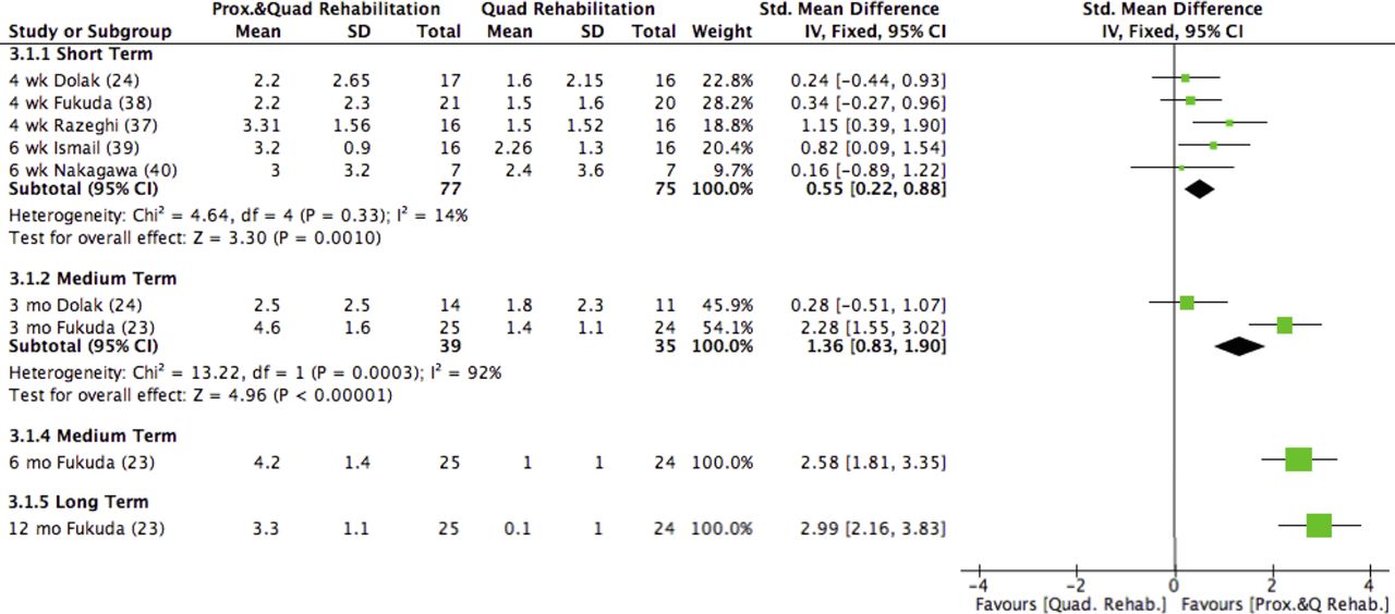
Four HQ23 ,38–40 and three LQ24 ,37 ,41 studies compared proximal combined with quadriceps rehabilitation to quadriceps rehabilitation alone (see online supplementary file 4 for further programme details), covering short,23 ,24 ,37 ,39–41 ,46 medium,23 ,24 and longer23 follow-up (figures 4 and 5).

Proximal and quadriceps rehabilitation compared with quadriceps rehabilitation for pain (‘short term’, <3 months; ‘medium term’, 3–12 months; ‘longer term’, ≥12 months; IV, inverse variance; Std., standard mean difference).

Proximal and quadriceps rehabilitation compared with quadriceps rehabilitation for function (LEFS, Lower Extremity Functional Score; AKPS, Anterior Knee Pain Score; ‘short term’, <3 months; ‘medium term’, 3–12 months; ‘longer term’, ≥12 months; IV, inverse variance; Std., standard mean difference).
Pain
Strong evidence (3 HQ38–40 and 2 LQ studies24 ,37) of a small effect indicated greater pain reduction favouring proximal combined with quadriceps rehabilitation using both OKC and CKC exercises compared to a quadriceps rehabilitation alone in the short term (I2=14%, p=0.33; SMD, 95% CI 0.55, 0.22 to 0.88). In the medium term, there was moderate evidence (1 HQ23 and 1 LQ study24) of a large effect indicating greater pain reduction following proximal combined with quadriceps rehabilitation using OKC and CKC compared to quadriceps rehabilitation alone in the medium term (I2=92%, p=0.0003; SMD, 95% CI 1.36, 0.83 to 1.90). Within the same HQ study, at 6 months, there was limited evidence (1 HQ study23) of a large effect indicating greater reduction in pain following proximal combined with quadriceps rehabilitation compared to quadriceps rehabilitation alone (SMD, 95% CI 2.58, 1.81 to 3.35).
In the longer term, there was limited evidence (1 HQ study23) of a large effect indicating greater pain reduction following proximal combined with quadriceps rehabilitation compared to quadriceps rehabilitation alone (SMD, 95% CI 2.99, 2.16 to 3.83).
Function
Strong evidence (2 HQ38 ,39 and 1 LQ24 study) of a small effect indicated greater functional patient-reported improvement (how measured) following proximal combined with quadriceps rehabilitation compared to quadriceps rehabilitation alone in the short term (I2=18%, p=0.30; SMD, 95% CI 0.42, 0.03 to 0.81). Limited evidence (1 HQ study38) indicated no difference in functional performance measured with the single leg hop test scores (SMD, 95% CI 0.32, −0.30 to 0.93) in the short term.
In the medium term, moderate evidence (1 HQ23 and 1 LQ24 study) of a large effect indicated increased patient reported function, measured by LEFS (I2=96%, p<0.00001; SMD, 95% CI 1.32, 0.75 to 1.89). Limited evidence (1 HQ study23) of a large effect indicated increased patient-reported function measured by AKPS (SMD, 95% CI 1.86, 1.18 to 2.54). Limited evidence (1 HQ study23) of large effect indicates increased performance-based function, measured by single leg hop scores (SMD, 95% CI 1.54, 0.89 to 2.18). Within the same study, at 6 months, limited evidence (1 HQ study23) of a large effect indicated increased patient-reported function, as measured by LEFS (SMD, 95% CI 2.49, 1.73 to 3.25), AKPS (SMD, 95% CI 1.86, 1.18 to 2.54), and performance-based function measured as single leg hop scores (SMD, 95% CI 1.85, 1.17 to 2.52).
In the longer term, limited evidence (1 HQ study23) of a large effect indicated greater patient-reported functional improvement as measured by LEFS (SMD, 95% CI 2.65, 1.86 to 3.43), AKPS (SMD, 95% CI 1.76, 1.09 to 2.42), and performance-based function measured with the single leg hop (SMD, 95% CI 2.06, 1.36 to 2.77) at 12 months, with proximal and quadriceps rehabilitation compared to quadriceps rehabilitation alone.
Mechanisms of proximal rehabilitation
Ten24–26 ,37 ,39 ,40 ,42 ,44–46 of the 14 studies explored variables with potential to explain proximal rehabilitation effects in the short term. No studies investigated mechanisms of effect at medium or longer-term follow-up.
Proximal rehabilitation—compared with—control
Limited evidence (2 LQ study26 ,46) of a large effect indicated that proximal rehabilitation using OKC band exercises increased isometric hip abduction strength (I2=81%, p<0.00001; SMD, 95% CI 1.69, 1.03 to 2.36) (figure 6). Very limited evidence (1 LQ study26) of a large effect indicated increased isometric hip external rotation strength in the OKC rehabilitation group in both the left (SMD, 95% CI 2.45, 1.43 to 3.46) and right (SMD, 95% CI 2.73, 1.66 to 3.80) hip. Very limited evidence (1 LQ study46) of a large effect indicated OKC proximal rehabilitation reduced the degree of knee valgum variability during consecutive footfalls while running (SMD, 95% CI 2.68, 1.54 to 3.82), but did not change peak knee genu valgum angles while running (SMD, 95% CI 0.83, −0.01 to 1.67).

Proximal rehabilitation compared with control group for strength and biomechanics in the short term (<3 months) (LHER, left proximal external rotation; RHER, right proximal external rotation; IV, inverse variance; Std., standard mean difference).
Proximal rehabilitation—compared with—quadriceps rehabilitation
Moderate evidence (1 HQ and 1 LQ study24 ,42) indicated no difference in maximal isometric strength for hip abduction (I2=60%, p=0.11; SMD, 95% CI 0.18, −0.08 to 0.44), external rotation (I2=0%, p=0.44; SMD, 95% CI 0.11, −0.15 to 0.37) or knee extension (I2=0%, p=0.33; SMD, 95% CI 0.09, −0.17 to 0.35) following a proximal (OKC) rehabilitation programme, as compared to a quadriceps (OKC/CKC) rehabilitation programme (figure 7). Limited evidence (1 HQ study42) indicated no difference in hip extension (SMD, 95% CI 0.09, −0.19 to 0.37) or internal rotation (SMD, 95% CI 0.02, −0.26 to 0.30) strength when comparing an OKC proximal with a CKC quadriceps rehabilitation programme.
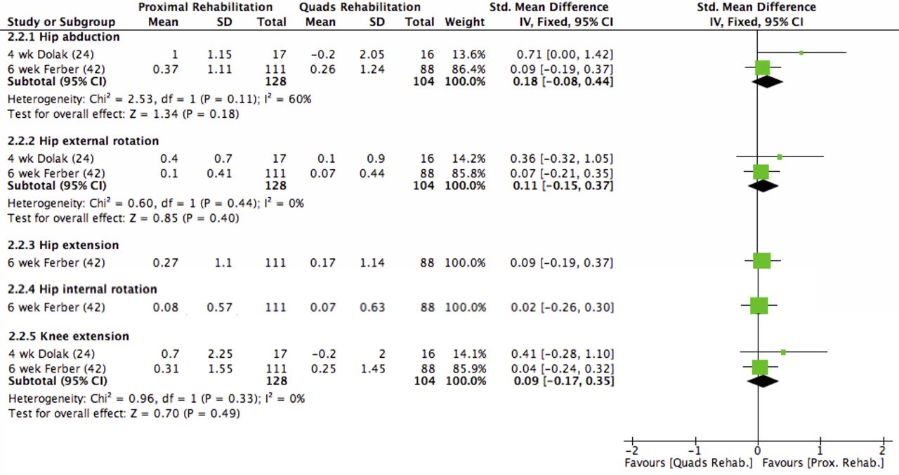
Proximal rehabilitation compared with quadriceps rehabilitation for isometric strength in the short term (<3 months) (IV, inverse variance; Std., standard mean difference).
Limited evidence (1 HQ study44) indicated proximal rehabilitation using CKC exercises, when compared with a CKC quadriceps rehabilitation protocol, increased the degree of pelvis anteversion (SMD, 95% CI 1.21, 0.43 to 1.98), hip flexion (SMD, 95% CI 1.16, 0.39 to 1.92), trunk contralateral inclination (SMD, 95% CI 0.90, 0.16 to 1.65), pelvis elevation (SMD, 95% CI 0.94, 0.19 to 1.68), hip abduction (SMD, 95% CI 2.20, 1.28 to 3.11), and knee adduction (SMD, 95% CI 0.81, 0.07 to 1.55) during a single leg squatting task. Additionally, anterior (SMD, 95% CI 2.03, 1.14 to 2.92), lateral (SMD, 95% CI 2.50, 1.53 to 3.46), and posterior (SMD, 95% CI 1.54, 0.73 to 2.36) trunk endurance (seconds), and proximal abductor (SMD, 95% CI 1.24, 0.47 to 2.02) and knee extensor (SMD, 95% CI 0.97, 0.22 to 1.72) torque (nm/kg) increased in the proximal rehabilitation group when compared with quadriceps rehabilitation.44
Proximal combined with quadriceps rehabilitation—compared with—quadriceps rehabilitation alone
Strong evidence (2 HQ studies39 ,40) indicated no difference in isokinetic, concentric and eccentric hip abduction, and external rotation strength following a proximal (OKC) combined with quadriceps (CKC) rehabilitation programme when compared to a quadriceps rehabilitation (CKC) programme alone (figure 8). Very limited evidence (1 LQ study24) indicated no difference in isometric hip abduction (SMD, 95% CI 0.54, −0.16 to 1.23) or external rotation (SMD, 95% CI 0.63, −0.07 to 1.33) strength following a proximal combined with quadriceps rehabilitation programme compared to quadriceps rehabilitation alone. Moderate evidence (2 LQ studies24 ,37) indicated no difference in isometric knee extension strength (I2=0%, p=0.37; SMD, 95% CI 0.07, −0.42 to 0.56) following comparison of proximal and quadriceps rehabilitation with quadriceps rehabilitation alone.

Proximal and quadriceps rehabilitation compared with quadriceps rehabilitation for strength in the short term (<3 months) (Abd, abduction; ER, external rotation; Ecc, eccentric; Ext, extension; HABD, hip abduction; HER, hip external rotation; IV, Inverse variance; Std., standard mean difference.
Proximal rehabilitation alone
One LQ study45 divided their cohort into ‘successful’ (≥1.5 cm reduction in VAS) and ‘unsuccessful’ groups, reporting an improvement in isometric proximal abduction and adduction strength was unrelated to success. Very limited evidence (1 LQ study25) indicated an improvement in lateral core endurance (p=0.001), hip abduction (p=0.008) and external rotation (p=0.03) isometric strength, and knee abduction internal moments (p=0.05) after an 8-week OKC and CKC neuromuscular activation intervention directed at the proximal musculature. A trend in increased anterior (p=0.06) and posterior (p=0.1) core endurance, and hip abduction moment (p=0.06) was reported in the same study.25 However, no change in rear foot eversion, knee abduction, hip adduction and internal rotation angles, rear foot inversion or hip external rotation moments were reported during a running task.25
Discussion
This systematic review and meta-analysis evaluated the effects of proximal muscle rehabilitation on pain and function in individuals with PFP and the potential mechanisms for effectiveness. Fourteen studies of varying quality were identified, including 11 RCTs.23 ,24 ,26 ,37–44 In the short term, strong evidence indicates proximal combined with quadriceps rehabilitation is significantly better at reducing pain than quadriceps rehabilitation alone,23 ,24 ,37 ,39–41 ,46 moderate evidence indicates proximal rehabilitation is better at improving pain compared to quadriceps rehabilitation alone,24 ,42–44 and very limited evidence indicates proximal rehabilitation reduces pain compared to a no intervention control.26 In the medium term, strong and moderate evidence indicates proximal and proximal combined with quadriceps rehabilitation, respectively, is more effective at reducing pain then quadriceps rehabilitation alone.23 ,43 ,44 In the longer term, limited evidence indicates proximal combined with quadriceps rehabilitation is more effective at reducing pain than quadriceps rehabilitation alone.23 Greater improvements in function were also reported for proximal, and proximal combined with quadriceps rehabilitation compared with quadriceps rehabilitation alone in the short (strong evidence), medium (strong to moderate evidence) and longer (limited evidence) term.23 ,24 ,38 ,39 Put together, these findings support the implementation of proximal muscle rehabilitation programmes for the management of PFP in clinical practice.
Effects of proximal rehabilitation
Pain
Strong evidence suggests that a combined proximal and quadriceps rehabilitation protocol using both OKC and CKC exercises results in superior short term outcomes of pain reduction when compared to CKC quadriceps rehabilitation alone. This pooled result is, however, driven by one LQ study37 that reported significant improvement in pain. In contrast, three24 ,38 ,40 of the five pooled studies reported no greater short-term pain reduction when compared to a quadriceps rehabilitation programme. An important consideration when interpreting these findings is that the CKC quadriceps exercises adopted within the three equivocal studies24 ,38 ,40 would also facilitate activation of proximal musculature and as such, could also be considered a combined proximal and quadriceps intervention. A similar story emerges when comparing proximal and quadriceps rehabilitation. Specifically, within a pooled group of heterogeneous studies that evaluated proximal compared with quadriceps rehabilitation, a large multicentre trial that compared an OKC proximal to a CKC quadriceps protocol demonstrated no significant differences in pain reduction between groups, but reported significant pain reduction within both groups in the short term.42 Adding proximal rehabilitation to quadriceps appears to offer the most favourable short-term outcome; however, it is clear that further research to identify the most effective rehabilitation programme design is required to maximise effectiveness.
In the medium term, pooled results indicate proximal rehabilitation more effectively reduced pain when compared to quadriceps rehabilitation alone. Among the pooled studies, exercise programme design differed significantly, with one utilising exclusively neuromuscular OKC43 and the other using neuromuscular and strength CKC44 exercises. Of these protocols, the neuromuscular OKC protocol resulted in the greatest positive symptom change, but further research directly comparing these different protocols is needed. Proximal combined with quadriceps rehabilitation was more effective than quadriceps rehabilitation in reducing pain in the medium and longer term, with treatment effects of large magnitude. The single study reporting significant pain reduction in the long-term used a combination of both OKC and CKC at an intensity assessed to be sufficient to evoke strength changes (ie, >70% 1 RM).23 These exercise parameters may be relevant to the successful outcome given that OKC exercises are commonly used clinically to allow for specific isolation of proximal musculature48 while attempting to reduce loading of the PFJ. These preliminary findings require further research to determine the most efficacious protocol to reduce pain, in the medium and long term.
Function
Fewer studies evaluated the effects of proximal muscle rehabilitation on functional outcomes, which limits the conclusions that can be drawn. Greater improvements in self-reported measures (WOMAC, LEFS, AKPS) and performance-based measures (Single Leg hop test) of function were observed in the short term following proximal rehabilitation compared to a no intervention control; and following proximal combined with quadriceps rehabilitation compared to quadriceps rehabilitation. Moderate evidence indicated no difference in function when comparing proximal with quadriceps rehabilitation, which differs from findings related to pain. It is possible that interventions that address the deficits in gluteal strength20 ,19 and activation21 and quadriceps strength19 that are evident in individuals with PFP could be sufficient to result in the short-term improvement of function, but not in the medium and longer term. Importantly, greater functional improvements are provided by proximal compared to quadriceps rehabilitation (strong evidence), and a combined proximal and quadriceps rehabilitation compared to quadriceps rehabilitation alone (limited to moderate evidence) in the medium and longer term. It is possible that superior pain outcomes with proximal compared to quadriceps rehabilitation in the short-term results from the quadriceps loading causing irritation to the PFJ, although further research documenting pain levels during exercise is needed to clarify this. These findings suggest that incorporating proximal rehabilitation into the management of PFP is beneficial to functional outcomes. However, considering the currently limited to moderate supporting evidence, further high quality studies are needed to confirm these positive results.
Mechanisms of proximal rehabilitation
This review evaluated potential short-term mechanisms of treatment effect, with studies reporting improvement in isometric24 ,26 ,46 and isokinetic strength,39 ,40 eccentric torque,44 trunk muscle endurance,44 single leg squat kinematics,44 reduced knee joint motion variability, 46 and reduced peak knee abduction internal moments.25 Changes in strength were measured in 10 of the 14 included studies. Limited evidence of large effect indicated greater improvement in isometric hip abduction strength in the short-term following a neuromuscular or strength-endurance resistance band intervention when compared to no intervention, offering a potential mechanism for changes in pain and function.26 ,46 However, given the absence of significant difference in maximal isometric hip abduction, extension, external and internal rotation, and knee extension strength when comparing OKC proximal rehabilitation to CKC quadriceps rehabilitation,42 it is also plausible that the positive effects of a rehabilitation intervention are not exclusively derived through changes in strength, but through a combination of more global lower limb strength changes, a change in lower limb biomechanics, possible central or systemic mechanisms,49 or most likely a combination of these factors.
Three studies exploring kinematic and/or kinetic change resulting from proximal rehabilitation reported small but significant changes about the hip and knee25 ,44 ,46 that offer potential mechanisms for treatment effects. Increased hip adduction and internal rotation has been identified previously as a risk factor for and is associated with PFP symptoms.5 ,14 Consequently interventions that demonstrate capacity to modify these parameters warrant further investigation. Although some evidence reports clinical measures are able to identify individuals with movement deficits and detect biomechanical change of small magnitude,50 it is important to ensure clinical tools are sufficiently sensitive to direct intervention choice and determine treatment effects.
Further exploration of the mechanisms for treatment effects following a rehabilitation intervention on muscle structure, neural innervation, systemic systems and biomechanics is needed to guide a tailored exercise approach to address patient-specific deficits and predict outcomes in individuals with PFP.
Clinical implications
Interventions that aim to improve strength, strength-endurance and neuromuscular activity of proximal musculature are effective in the management of PFP, and should be incorporated in clinical practice. From within the largest high quality study that explored the effect of proximal compared with quadriceps rehabilitation, OKC proximal rehabilitation in isolation was not more effective at reducing pain than a CKC quadriceps rehabilitation programme42 in the short term. These findings suggest CKC quadriceps rehabilitation is as effective at increasing proximal strength as an isolated OKC proximal rehabilitation programme and have potentially important clinical implications for exercise intervention design. These suggest that clinicians could choose different treatment approaches and achieve the same or similar strength gains. Subsequently, this would allow clinicians to be guided by patient response, preference or available equipment, without negatively impacting on patient care.
Proximal rehabilitation combined with quadriceps rehabilitation using both OKC and CKC exercises produced better pain and functional outcomes in the short,44 medium and longer term23 compared to quadriceps rehabilitation alone. Therefore, therapists treating PFP should aim to prescribe exercise interventions targeting the quadriceps and proximal musculature in individuals with PFP using a combination of OKC and CKC exercises.
Rehabilitation exercises from studies included in this review were completed between 3 and 7 times per week, with the intensity of the programmes varying significantly. Exercise frequency did not appear to be dependent on the type and intensity of the rehabilitation protocols prescribed. Given established guidelines33 that indicate neuromuscular training has greater effect if performed frequently (daily) and strength training less frequently (2–3×per week) on physiological adaptation, the specificity of exercise frequency in the management of PFP is lacking within the current evidence base and requires further research.
Given that the primary goal of PFP treatment is often pain reduction, utilising exercise parameters that do not aggravate symptoms is important. Consequently, utilising OKC exercises in the short term or CKC exercises within finite pain limits throughout the rehabilitation process is advocated by the current evidence.23 ,24 ,44 It is unclear at present whether these parameters are essential for a successful outcome; further research is required to determine the most effective protocol design. Furthermore, identification of individuals who are more likely to respond to a specific proximal intervention approach is an important consideration given the dearth of evidence exploring indicators for proximal intervention success.51
Limitations
There are limitations that need to be considered when interpreting the results of this review. Variability in study design, type of protocol (OKC or CKC), and differing outcome measures limited further data pooling. Where pooling was possible, heterogeneity of rehabilitative exercise prescription remained evident in both exercise frequency and intensity. Nonetheless, given the paucity of available evidence, we felt that data pooling was valuable to strengthening the findings of the review. Non-English language publications were not sought in this review. While it is arguable that high-quality RCTs would aim to be published in higher impact journals written in the English language, identification of further trial data may have influenced the outcomes of analysis. Assessment of methodological quality was completed using the PEDro scale. It was identified that only 43% of the included 11 studies blinded the assessor to the intervention delivered and only one study attempted to blind the participants to their group allocation. Given growing evidence for proximal intervention efficacy, future studies should aim to blind the participants and researchers to the allocated group. Finally, it has been identified that mechanobiological determinants of exercise prescription are poorly reported or defined within the included rehabilitation studies. To ensure clinical utility and consistency of exercise parameter reporting within future studies, these determinants must be detailed.
Conclusion
The best available evidence indicates that proximal rehabilitation with or without simultaneous quadriceps rehabilitation is beneficial to pain and function in individuals with PFP in the short and medium term. While fewer studies have evaluated long-term effects, the limited evidence available indicates proximal and quadriceps rehabilitation combined has greater positive benefit on pain and function than quadriceps rehabilitation alone. A combination of both OKC and CKC exercise are most likely to result in favourable outcomes. Given variability in rehabilitation protocols within the current literature, further studies designed to identify the most effective protocol by considering exercise type, load and dose are required. To improve clinical applicability, these studies must detail specific exercise descriptors. Regardless, proximal rehabilitation should be incorporated into clinical reasoning paradigms for the management of PFP.
Summary box
Proximal combined with quadriceps rehabilitation reduces pain and improves function more than quadriceps rehabilitation in the short, medium and long term.
Proximal rehabilitation is more effective than quadriceps rehabilitation in decreasing pain in the short term, and decreasing pain and improving function in the medium term.
Maximum hip muscle isometric strength can be increased by proximal rehabilitation compared to no exercise controls, but not significantly more than a closed kinetic chain quadriceps focused rehabilitation programme.
References
Supplementary materials
Supplementary Data
This web only file has been produced by the BMJ Publishing Group from an electronic file supplied by the author(s) and has not been edited for content.
- Data supplement 1 - Online supplement
- Data supplement 2 - Online supplement
- Data supplement 3 - Online supplement
- Data supplement 4 - Online supplement
- Data supplement 5 - Online supplement
- Data supplement 6 - Online supplement
Footnotes
Funding #TEAM_PFP would like to acknowledge the Private Physiotherapy Education Fund (PPEF) for funding some of SL's time to complete this review. DM is part funded by the NIHR/HEE Senior Clinical Lecturer scheme. This article presents independent research part funded by the National Institute for Health Research (NIHR). The views expressed are those of the authors and not necessarily those of the NHS, the NIHR or the Department of Health.
Competing interests None declared.
Provenance and peer review Not commissioned; externally peer reviewed.