Article Text

Abstract
Background Cam morphology, a distinct bony morphology of the hip, is prevalent in many athletes, and a risk factor for hip-related pain and osteoarthritis. Secondary cam morphology, due to existing or previous hip disease (eg, Legg-Calve-Perthes disease), is well-described. Cam morphology not clearly associated with a disease is a challenging concept for clinicians, scientists and patients. We propose this morphology, which likely develops during skeletal maturation as a physiological response to load, should be referred to as primary cam morphology. The aim of this study was to introduce and clarify the concept of primary cam morphology.
Design We conducted a concept analysis of primary cam morphology using articles that reported risk factors associated with primary cam morphology; we excluded articles on secondary cam morphology. The concept analysis method is a rigorous eight-step process designed to clarify complex ‘concepts’; the end product is a precise definition that supports the theoretical basis of the chosen concept.
Results We propose five defining attributes of primary cam morphology—tissue type, size, site, shape and ownership—in a new conceptual and operational definition. Primary cam morphology is a cartilage or bony prominence (bump) of varying size at the femoral head-neck junction, which changes the shape of the femoral head from spherical to aspherical. It often occurs in asymptomatic male athletes in both hips. The cartilage or bone alpha angle (calculated from radiographs, CT or MRI) is the most common method to measure cam morphology. We found inconsistent reporting of primary cam morphology taxonomy, terminology, and how the morphology is operationalised.
Conclusion We introduce and clarify primary cam morphology, and propose a new conceptual and operational definition. Several elements of the concept of primary cam morphology remain unclear and contested. Experts need to agree on the new taxonomy, terminology and definition that better reflect the primary cam morphology landscape—a bog-standard bump in most athletic hips, and a possible hip disease burden in a selected few.
- hip
- athletes
- adolescent
- training load
- sporting injuries
Data availability statement
All data relevant to the study are included in the article or uploaded as supplementary information.
This is an open access article distributed in accordance with the Creative Commons Attribution Non Commercial (CC BY-NC 4.0) license, which permits others to distribute, remix, adapt, build upon this work non-commercially, and license their derivative works on different terms, provided the original work is properly cited, appropriate credit is given, any changes made indicated, and the use is non-commercial. See: http://creativecommons.org/licenses/by-nc/4.0/.
Statistics from Altmetric.com
Introduction
Femoroacetabular impingement (FAI) syndrome and hip osteoarthritis (OA) are common causes of hip-related pain and strongly associated with cam morphology of the hip.1–5 Secondary cam morphology, due to pre-existing hip disease or acute trauma including Perthes disease, slipped capital femoral epiphysis, healed proximal femoral fractures or acute fracture, is well-described.3 5 Cam morphology not associated with a primary disease is a challenging concept for clinicians, scientists and patients. We propose this morphology, which likely develops during skeletal maturation as a physiological response to skeletal loading patterns at the hip, should be referred to as primary cam morphology.
A primary medical condition is one that arises spontaneously and is not associated with, or caused by a previous disease, injury or acute event.6 For example, primary osteoporosis, bone loss due to ageing or the loss of sex steroids at menopause, differs from secondary osteoporosis which is due to conditions such as thyroid hormone imbalance or renal disease.7 8 Thus, primary cam morphology is cam morphology that is not caused by previous disease, injury or an acute event.
This study is laser-focussed on primary cam morphology because we feel that the community of sports medicine clinicians, researchers and patients interested in hip-related pain needs to clearly delineate what they mean when using terms such as ‘cam morphology’, ‘cam lesions’, ‘cam-type impingement’ or ‘cam deformity’.9–11 Clarifying the taxonomy, terminology and definition of primary cam morphology are key steps to assist the community to distinguish between a normal variant (‘bog-standard’) and a pathology (‘burden’) in athletes with primary cam morphology.
The aim of this study was to introduce and clarify the concept of primary cam morphology using formal method of ‘concept analysis’ by Walker and Avant. Specifically, we aimed to:
describe the taxonomy and classification of primary cam morphology;
synthesise how terminology is currently used;
list the defining attributes of primary cam morphology and how they are operationalised (their ‘empirical referents’);
identify the antecedents and consequences of primary cam morphology;
propose a conceptual and operational definition for primary cam morphology.
Methods
Concept analysis
The concept analysis method by Walker and Avant is a rigorous eight-step process to examine the basic elements of a concept.12 The results are precise conceptual and operational definitions, and clear communication as a basis for research and clinical practice.12 Concept analysis has been used in other healthcare disciplines to clarify the characteristics and attributes of abstract concepts such as chronic fatigue.12–15 Concept analysis has not been previously applied in the field of sports and exercise medicine and may establish a foundation for higher quality research and clinical decision making.12
Concept analysis method is an intellectual exercise and a strategy to construct theory; it is not a mere summary of the concept. Concept analysis guides a discipline and links research, theory and practice by providing clear conceptual foundations. Without these foundations, research quality and theory construction of any discipline is weakened and its maturity compromised.12 16 We discuss concept analysis purposes, processes, examples and relevant terminology in online supplemental material folder 1: figure 1; tables 1 and 2.
Supplemental material
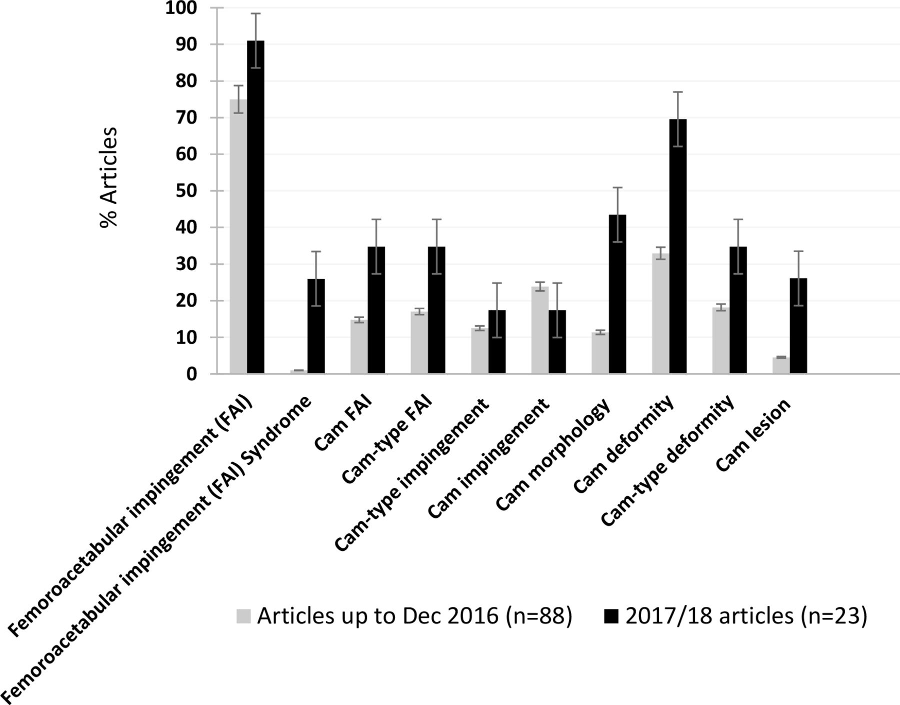
Time trend for the 10 most-used terms relating to primary cam morphology—all articles from 2016 and earlier (grey bars, n=88) compared with 2017/18 articles (dark bars, n=23); many articles used more than one term. Error bars=SEs.
Primary cam morphology, its defining attributes and empirical referents; quotes from articles in italics (refer to online supplemental material folder 3 for more detail)
Supplemental material
Primary cam morphology conceptual definition statements based on its attributes, empirical referents, antecedents and consequences
We applied the eight-step concept analysis process to the concept of primary cam morphology.
Step 1: select a concept
Consensus statements from leading experts on hip-related pain, the Warwick consensus statement on FAI syndrome4 and the International Hip-related Pain Research Network consensus on the classification, definition and diagnostic criteria on hip-related pain in young and middle-aged active adults,17 both recommended further research on cam morphology. The authors of this manuscript are all members of the Young Athlete’s Hip Research (YAHiR) Collaboration, an international grouping of multiprofession stakeholders whose aim is to increase value and reduce waste through higher quality research on the aetiology, treatment and prognosis of conditions that affect the young person’s hip (including bony morphologies). ‘Yahir’ is an Arabic name and means ‘they will enlighten’; the YAHiR Collaboration aims to ‘enlighten’ better clinical decisions for patients through higher quality research.
We selected primary cam morphology as a distinct and important concept for clinical practice, education and research. It is important to distinguish between primary and secondary cam morphology because, although they are related concepts, they have distinctly different aetiology and clinical management. Primary cam morphology likely develops during maturation as a physiological response to specific, but to date unclear physical loading patterns, and is therefore important for many athletic cohorts. Clarity on primary cam morphology taxonomy, and conceptual and operational definitions will be key for future long-term research on its aetiology, clinical management and prognosis.
Step 2: determine the aims and purpose of the analysis
Members of the YAHiR Collaboration agreed that primary cam morphology was a distinct, important concept; there was a need to clarify the concept to permit more rigorous and evidence-based research on primary cam morphology. The aim of this study was, therefore, to perform an in-depth concept analysis of the concept of primary cam morphology. We describe its taxonomy, synthesise how terminology is currently being used, list the defining attributes, identify its antecedents and consequences and propose a conceptual and operational definition for primary cam morphology.
It will help clinicians, scientists and patients to better understand and manage hip conditions related to primary cam morphology in athletes, including hip-related pain, FAI syndrome and OA of the hip.
Step 3: identify all the uses of the concept and select the literature
The concept of cam morphology is normally used in the context of bony morphologies of the hip, FAI and FAI syndrome and OA of the hip. Any risk factor study (aetiological or prognostic) relies on clear conceptual and operational definitions for the specific condition/disease to avoid, among other biases, misclassification bias. The scope of this study is to introduce and understand primary cam morphology—cam morphology that develops spontaneously, likely as a normal physiological response to load—in the context of its risk factors. We used the studies identified for a separate ongoing systematic review of risk factors for primary cam morphology. We provide the systematic review methods as supplementary material (online supplemental material folder 2): study eligibility criteria, search strategy, study selection, data extraction (domains adapted from the CHecklist for critical Appraisal and data extraction for Systematic Reviews of prediction Modelling Studies (CHARMS18), quality and risk of bias assessment (combining the Quality in Prognosis Studies tool19 20 and Risk of Bias tool for Non-randomised Studies21) and data synthesis and analysis. The systematic review protocol is available online: bit.ly/cammorph.22
Supplemental material
Step 4: determine the defining attributes
We extracted primary cam morphology conceptual definitions (how authors conceptually defined cam morphology) and operational definitions (how the different attributes were measured) from the studies included in the review. We then took a systematic and purposeful approach to discover the defining conceptual and operational attributes, antecedents and consequences. We did this by:(1) reading the included articles (HPD read all the included articles and three coauthors (CLA, AS, AW) each read one-third of them), (2) identifying and extracting the different conceptual and operational characteristics of primary cam morphology: (HPD developed the initial conceptual and operational framework, antecedents and consequences, and refined this with the coauthors CLA, AS, AW, ABM, SMcA and SG-J), (3) placing the frequently occurring characteristics into a coding scheme (HPD did this using Atlas.ti software), (4) grouping the characteristics and classifying them into categories and subcategories, (5) discussing the categories and subcategories, and underlying characteristics in the author team and with other experts, (6) renaming the categories as attributes, (7) randomly assigning two papers to coauthors (AS, SMcA and ABM) for coding using the attribute framework and Excel and (8) further refining the attribute framework after coauthor coding and feedback.
We present examples from included studies to explain each attribute as part of the results.
Step 5: identify a model case
HPD (in collaboration with the coauthor team) identified a model case based on real-life experiences working with patients with primary cam morphology and/or FAI syndrome. We refined and developed this case as a narrative to illustrate conceptual and operational definitions for primary cam morphology.
Step 6: identify additional cases
We wrote corresponding narratives for additional borderline and contrary cases to further illustrate the concept of primary cam morphology. Additional cases describe borderline cases, related cases, contrary cases and invented cases. This is an important step as it may be difficult to determine the defining attributes that most closely represent primary cam morphology. We therefore describe additional cases to help refine the best-fit defining attributes.12
Step 7: identify antecedents and consequences
Antecedents and consequences illuminate a concept’s context. According to Walker and Avant,12 a defining attribute cannot be either an antecedent or a consequence. Antecedents are events that must arise or be in place prior to a concept’s occurrence. For instance, if a tibial stress fracture is the concept under investigation, an antecedent could be prior high-volume training on a hard surface. The consequences are events or incidents that can arise as a result of the concept. Chronic non-union might be a consequence of an anterior cortical tibial stress fracture. The antecedents and consequences of primary cam morphology were extracted from risk factor papers and discussed among authors (who have extensive clinical and research experience in the field). Antecedents and consequences serve to refine the defining attributes. All the authors of this manuscript discussed the important antecedents and consequences related to primary cam morphology and reached consensus.
Step 8: define empirical referents
We described how various authors observed and measured the different conceptual attributes for primary cam morphology, which could relate to patient history, clinical examination and/or imaging investigations (empirical referents—“the means by which you can recognise or measure the defining characteristics or attributes” of a concept12) (online supplemental material folder 1 further clarifies the term: ‘empirical referent’).
Results
We present the results according to the above eight steps, as described by Walker and Avant, combining steps 1–3 on the literature used for the concept analysis, and steps 5 and 6 on model and additional cases.12
Steps 1–3: select the concept; determine the aims and purpose of the analysis; identify all the uses of the concept and select the literature
Primary cam morphology is a distinct and important concept for clinical practice and research. Our initial database search yielded 10 519 records, of which 111 met the risk factor systematic review eligibility criteria. We included all 111 articles in this concept analysis.
The concept: primary cam morphology taxonomy and terminology
There were 206 different terms related to cam morphology in the 111 included articles, which can be divided into three categories: (1) cam morphology as it relates to FAI, (2) ‘morphology’ and its related terms and (3) ‘lesion’, ‘deformity’, ‘abnormality’ and related terms. Most of the included articles referred to cam morphology in the context of FAI and FAI syndrome (78% and 6% of the 111 included articles). Cam FAI and cam-type FAI were used in 19% and 21% of the included articles, while 23% used cam impingement and 14% used cam-type impingement. Cam lesion, cam deformity and cam-type deformity were used in 9%, 41% and 22% of the included articles, respectively. Many articles use more than one term (some up to five) for the same concept.
We compared the most-used terms in all articles from 2016 and earlier (n=88) and those published in 2017/18 (n=23) (ie, articles published at least 2 months after the Warwick consensus paper recommended to use ‘cam morphology’ and avoid ‘lesion’ and ‘deformity’) (figure 1).4 There was greater use of ‘cam morphology’ in the 2017/18 articles compared with articles from 2016 and earlier (43% vs 11%), and also greater use of ‘FAI syndrome’ (26% vs 1%), ‘cam deformity’ (70% vs 33%), ‘cam lesion’ (26% vs 5%) and ‘cam FAI’ (35% vs 15%) (figure 1).
Step 4: attributes
We describe five attributes and combine step 8, empirical referents, with each attribute to describe how it was recognised or measured (operationalised) (table 1). Refer to online supplemental material folder 3 for more detail.
Attribute 1: tissue types—cartilage or bone
All but one of the included articles described cam morphology as a bony entity. The article describing cartilage and bony cam morphology used 3 T MRI to distinguish between cartilage and bone.23 They showed that the cartilage alpha angle increased as early as age 10 years, qualitatively representing soft-tissue hypertrophy at the head-neck junction, preceding extension of the ossified epiphysis. Cartilage alpha angle might therefore reflect the hip shape better than the secondary ossification centre in skeletally immature individuals. It is likely non-ossified structures that impact in FAI in these young hips but more research is needed to confirm this. Bony primary cam morphology is described and measured on radiographs, CT scans and MRI at the time of and after femoral head physeal closure.
Attribute 2: size
Primary cam morphology is a three-dimensional entity of variable size. It was described in the included articles as ‘small’, ‘moderate’, ‘large’, ‘pathological’, ‘significant’, ‘severe’ or ‘definite’. Assigning these categories can be a qualitative judgement (subjective impression of shape and size on imaging) or quantified on imaging through various measures such as the alpha angle (figures 2A and 3C).

(A) Increasing size of cam morphology (anterior cam morphology in a sagittal oblique or transverse plane; left hip; superior cam morphology in coronal (frontal) plane; left hip); 1=acetabulum; 2=femur head; 3=femur neck; 4=cam morphology. (B) Anterior cam morphology.

(A) Schematic representation of radial images around the axis of the femoral neck. The radial MRI planes are perpendicular to the femoral head-neck axis (sagittal oblique MRI localiser). (B) The radial cuts rotate clockwise in 30° intervals around the femoral head-neck axis. The coronal plane (12 o’clock) is parallel to the axis of the proximal femur diaphysis. Adapted from Reichenbach et al,32 Siebenrock et al, 95 Dudda et al.96
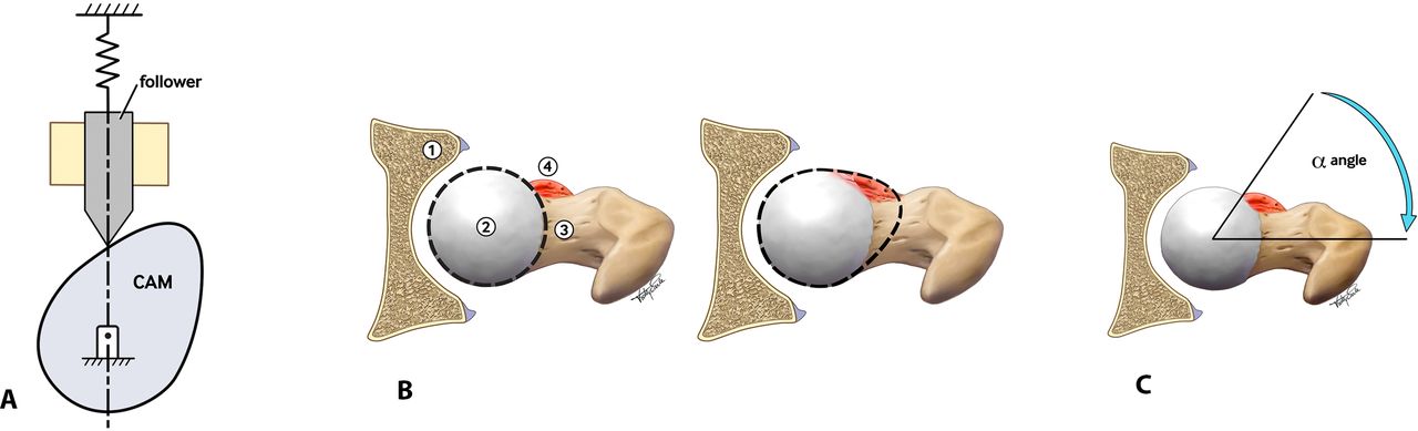
A cam disc/shaft: ‘cam’ is a rotating or sliding piece, such as an eccentric wheel or a cylinder with an irregular (oval) shape in a mechanical linkage used to transform rotary motion into linear motion or vice versa.40(B) Cam morphology—changing the shape of the femoral head from spherical to aspherical (this can be an anterior or superior position; left hip); 1=acetabulum; 2=femur head; 3=femur neck; 4=cam morphology. (C) The alpha angle: angle between a line joining the centre of the femoral head with the centre of the neck at its narrowest point, and a line from the centre of the femoral head to a point where the distance from the centre of the head exceeds its radius.97
Attribute 3: location (site)
Primary cam morphology location refers to the general anatomical area (femoral head-neck junction; attribute 3.1), and the specific anatomical location (attributes 3.2 and 3.3), depending on the type of imaging used to operationalise the morphology (two-dimensional or three-dimensional) (figures 2A,B, 4A,B).
Attribute 3.1: femoral head-neck junction
Primary cam morphology develops at the femoral head-neck junction. The location is also described as ‘proximal femur’,24–28 ‘femoral’, ‘femoral head’, ‘femoral head-neck transition’, ‘femoral head-neck type’.
Attribute 3.2: anterior, anterolateral, lateral, inferior, posterior, superior, anterior-superior, superior-anterior
Primary cam morphology is a three-dimensional entity usually located on the anterosuperior aspect of the femoral neck.29 Anterior and lateral primary cam morphology are visible and measured on true lateral-pelvis and AP-pelvis radiographs, respectively (two-dimensional imaging) while cam morphology in any femoral head-neck position is visible and measured on three-dimensional imaging (CT scan or MRI) (figure 4A). Several lateral views exist to visualise other parts of the head-neck junction (eg, corresponding to the anterolateral region). Despite being a two-dimensional image, radiographs can still capture the presence of cam morphology quite accurately. However, it does not always capture ‘peak’ cam morphology. The size and position of the bony prominence may vary. One paper suggests it is more superior in males and more anterior in females.30
Attribute 3.3: different o’clock positions of the femoral head (12 o’clock to 11 o’clock)
Many authors (≈40% of the total included articles) used a clock face system (figure 4A B) to describe the location of cam morphology on radial MRI or CT scan sequences around the axis of the femoral neck, normally 30° intervals with 12 o’clock as the superior location, and 3 o’clock, 6 o’clock and 9 o’clock as the anterior, inferior and posterior locations, respectively.23–26 31–37 The most frequent positions used are 12 o’clock to 3 o’clock.
Attribute 4: shape
The included articles used a variety of terms to describe or refer to the ‘cam shape’. These terms include ‘cam morphology’, ‘FAI morphology’, ‘morphological variation’, ‘pathomorphology’, ‘pistol-grip deformity’, ‘tilt deformity’, ‘bump’, ‘hump’, ‘prominence’, ‘reduced (less; diminished) femoral head-neck junction concavity’, ‘incongruity’, ‘convex’, ‘flattening’, ‘asphericity (aspherical; non-spherical)’ and ‘oval-shape’. The normal anatomy and morphology of the femoral head (caput femoris) and neck (collum femoris) are well documented.38 39 In Mechanical Engineering, ‘cam’ refers to an irregular aspherical rotating or sliding piece (figure 3A).40 Cam morphology in orthopaedics refers to an aspherical or cam-shaped femoral head (figure 3B).
Attribute 5: ownership
Primary cam morphology is more common in male athletes and occurs in one, but more often in both hips. It is reported per hip and/or per person in the included articles.
Attribute 5.1: sex/gender
Primary cam morphology is more prevalent in males compared with females.41 42 More research in female cohorts is needed.
Attribute 5.2: athletes
Primary cam morphology is more prevalent in athletes compared with non-athletes. There is strong evidence that high activity levels during adolescence promote cam morphology development with a dose-response relationship.23 31 43
Attribute 5.3: one or both hips (per hip; per person)
Some included articles analysed and reported cam morphology in both hips for each research participant as a dichotomous outcome variable (using a range of different cut-off values), a continuous outcome variable or both (table 1).
Some included articles analysed and reported cam morphology in one hip (‘per person’) for each research participant: either the ‘right or the left hip’, the ‘dominant hip’, the ‘kicking leg’, a ‘random hip’ or a ‘symptomatic hip’ (table 1).
Attribute 5.4: symptoms
The majority of individuals with primary cam morphology are symptom free. In a 2-year prospective cohort study of professional adult male football players, bony morphology, including cam morphology, was not associated with the risk of groin injuries. Despite the high prevalence of cam morphology (71% of players), only 1 of 113 index hip/groin injuries recorded was hip-related.44
Step 8: empirical referents
Primary cam morphology is only visible on imaging or during open or arthroscopic hip joint surgery. The included articles used imaging to observe or measure (operationalise) primary cam morphology, qualitatively (visual) and/or quantitatively (measuring a specific imaging outcome variable).
Imaging used for primary cam morphology
Primary cam morphology was measured on radiographs, dual-energy X-ray absorptiometry (DXA), CT scans and MRI.
Radiographs
The following radiographs were used to operationalise cam morphology applying a range of different outcome measures: AP pelvis, Dunn 45, Frog-leg lateral, cross-table lateral,45–48 Sugioka view,49 standing false profile hip27 and Von Rosen view,36 50 Lauenstein radiograph.51
Dual-energy X-ray absorptiometry
One of the included articles used posterior-anterior DXA bone mineral density images to quantify bony morphology of the hip.52
CT scan
CT scans were used in 18 of the 111 included articles to operationalise cam morphology.26 30 35 53–67 Ng et al 62 describe axial alpha angles measured on oblique-axial plane of the longitudinal femoral head-neck axis (cam deformity in the anterior aspect of the femoral head), and radial alpha angles obtained through a 1:30 clockface rotation about the longitudinal femoral head-neck axis (anterosuperior quadrant). Axial alpha angle >50.5° or radial alpha angle >60° were considered as cam deformity.62 Speirs et al measured the alpha angle on two images to evaluate the femoral head-neck junction anteriorly and anterosuperiorly in the traditional axial oblique (3:00) and radial 1:30 planes, respectively. The classified asymptomatic subjects with an alpha angle ≥50.5° measured in the 3:00 plane or >60° in the 1:30 plane as ‘bump’.64
Magnetic resonance imaging
MRI of different magnetic field strengths, 0.5 T, 1 T, 1.5 T and 3 T were used to operationalise cam morphology and the important associate structures (eg, physeal growth plate, labrum and joint cartilage). The authors describe coils, ‘body coil for signal transmission and a flexible four-channel surface coil for signal reception’,35 relaxation time: T1ρ, turbo spin echo (TSE)35 and planes: sagittal,23 sagittal-oblique,35 radial,23 68 ‘axial angled on the femoral neck’,33 axial-oblique,69 ‘transverse oblique with radial images reformatted by using the femoral neck long axis as a rotation axis’,70 ‘axial-oblique sagittal and coronal’ and71 coronal-oblique.72
Outcome measures
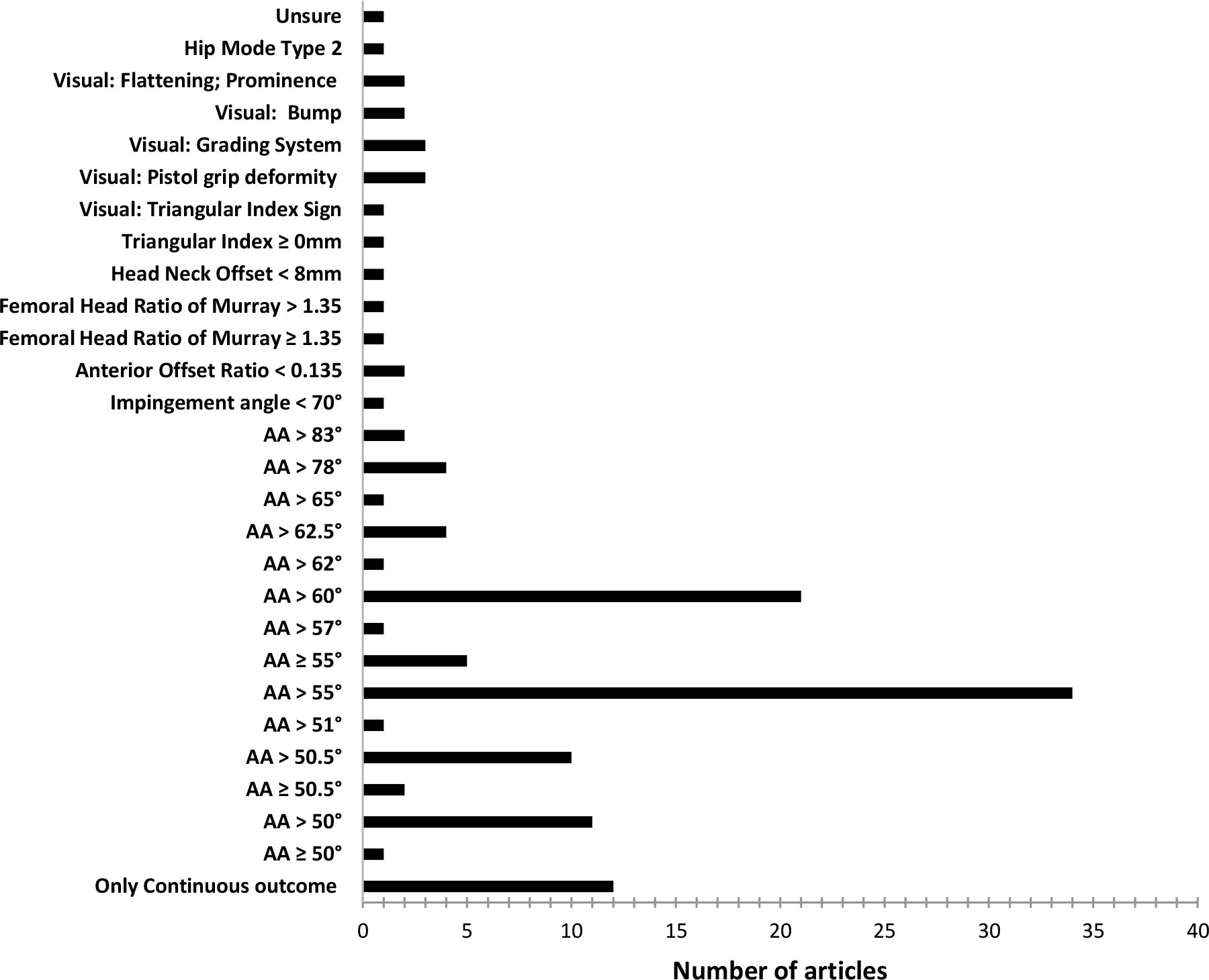
The included articles used different imaging outcome measures to report the shape and size of cam morphology (figure 5). These include alpha angle (degrees) (figure 3C), impingement angle (degrees), offset measure (mm), offset ratio, femoral head ratio of Murray, triangular index and the relationship between the width of the femoral neck and diameter of the femoral head.

Cam morphology outcome measures and prevalence definitions based on a dichotomous outcome measure. AA, alpha angle.
The included articles reported these outcome variables as continuous and/or dichotomous using different cut-off values. There was no consensus on an operational definition for cam morphology based on any of the outcome variables in the literature (figure 5).
Steps 5 and 6: model and additional cases
All the authors of this manuscript, many with extensive relevant clinical experience in the field, contributed to craft the model and additional cases to inform the concept. We describe a model case of primary cam morphology in a male football player aged 15 years. We wrote corresponding narratives for additional cases, describing borderline cases, related cases and contrary cases to further illustrate the concept of primary cam morphology. This is an important step as it may be difficult to determine the defining attributes that most closely represent primary cam morphology. We therefore describe three additional cases to help refine the best-fit defining attributes: (1) primary mixed morphology, (2) hip dysplasia and (3) secondary cam morphology due to slipped capital femoral epiphysis (online supplemental material folder 1).
Step 7: antecedents and consequences
The science concerning primary cam morphology, including its aetiology and prognosis, is not settled. No high-quality prospective studies with an adequate follow-up time exist on primary cam morphology aetiology or prognosis. This concept analysis will inform higher quality future research, including expert opinion and consensus agreement (or expert dissent for discussion) on taxonomy, terminology, definitions and imaging outcome measures. A collaborative approach to multicohort prospective aetiology and prognosis studies provides the opportunity to share higher quality, uniform research data.
Antecedents
We identified three primary cam morphology antecedents: (1) young adolescents with no other disorders of the hip (absence of conditions associated with secondary cam morphology), (2) an open femoral capital physis and (3) high shear-type load as the likely causative risk factor (volume and type of load are not well understood; probably external rotation with flexion leading to a combination of axial and rotational shear forces), and other unconfirmed risk factors (refer to online supplemental material folder 1 for more detail).
Consequences
Primary cam morphology can cause FAI, FAI syndrome, microscopic or macroscopic cartilage and/or labral damage and finally hip OA (refer to online supplemental material folder 1 for more detail).
Conceptual and operational definition for primary cam morphology
Based on the defining attributes and empirical referents for primary cam morphology, the clinical cases, antecedents and consequences, we propose the following conceptual and operational definition for primary cam morphology (table 2 and figure 6):
Primary cam morphology is a cartilage or bony prominence (bump) of varying size at any location around the femoral head-neck junction, which changes the shape of the femoral head from spherical to aspherical. It often occurs in asymptomatic male athletes in both hips. The most common outcome measure is a cartilage or bone alpha angle as a dichotomised or continuous variable on radiographs, CT scans or MRI, reported per hip, per person or both. Primary cam morphology likely develops during skeletal maturation in young adolescents (with no current or previous hip disease), as a normal physiological response to high-load sporting activity and other unconfirmed risk factors.

(A) Left hip in neutral position. (B) Left hip in internal rotation. Primary cam morphology is a cartilage or bony prominence (bump) of varying size at any location around the femoral head-neck junction of the hip, which changes the shape of the femoral head from spherical to aspherical. It often occurs in asymptomatic male athletes in both hips. The most common outcome measure is a cartilage or bone alpha angle as a dichotomised or continuous variable on radiographs, CT scans or MRI, and reported per hip, per person or both. Primary cam morphology likely develops during skeletal maturation in young adolescents (with no current or previous hip disease), as a normal physiological response to high-load sporting activity and other unconfirmed risk factors.
Discussion
In this first concept analysis of primary cam morphology in the disciplines of sports medicine, orthopaedics, radiology and physiotherapy, we introduce primary cam morphology and propose five defining attributes—tissue type, size, site, shape and ownership—in a new conceptual and operational definition. Concept analysis is a rigorous method to clarify a concept. Here, we highlight that taxonomy for the morphology is inconsistent, and terminology and how the morphology is described (imaging) are equally variable. We outline the clinically important findings related to primary cam morphology—a distinct concept in many (male) athletes, inconsequential (‘bog-standard’) in most, but an important risk factor for early hip disease in some.
Primary cam morphology taxonomy (classification) and terminology
Taxonomy: there is no agreed taxonomy for primary cam morphology. Primary cam morphology is an important concept—a normal physiological response to load, hence bog-standard in most athletes. Yet, in some athletes, this morphology can be associated with burdensome hip disease such as FAI syndrome and OA (online supplemental material folder 1: figure 2A and B). Disease taxonomy (naming, describing and classifying disease into domains and subcategories), underpins communication and research.73–76 The International Classification of Diseases (ICD) has no detailed taxonomy for FAI syndrome, a hip disease with described clinical and imaging characteristics, including cam morphology.4 77 The ICD-11 code, FA34.5, refers only to ‘impingement syndrome of the hip’ without detailing the associated bony morphology (online supplemental material folder 1: figure 2A and B).77 This vagueness is a problem for clinicians and researchers in sports medicine, orthopaedics, radiology and physiotherapy.
Primary cam morphology is more common in male athletes. An athlete with hip-related pain, who participated regularly in impact sports during maturation and has no previous hip disease, a positive Flexion Adduction Internal Rotation test and cam morphology on imaging, has FAI syndrome and primary cam morphology. Clinicians should reason differently when they manage these patients compared with a patient with FAI syndrome and secondary cam morphology.
Terminology: the terminology for cam morphology is only consistent in its inconsistency. The 2016 Warwick consensus on FAI syndrome, endorsed by 25 clinical societies, recommended the term ‘cam morphology’. The authors recommended eschewing terms such as ‘deformity’, ‘abnormality’ or ‘lesion’—to avoid attributing ‘pathology’ to an anatomical feature.4 Our concept analysis indicates that the Warwick nomenclature has not yet gone viral among cam morphology researchers. One reason might be that there is no consensus on terminology, definitions and imaging outcome measures specific to cam morphology—when anatomy flips to pathology. Overall, only 11% of the 88 articles from 2016 and earlier, included in our concept analysis data set, used the term ’cam morphology’, and this increased to 43% after 2016.
A pragmatic FAI and cam morphology taxonomy and terminology, to include primary and secondary cam morphology, provides a conceptual framework that will allow clinicians, researchers and patients to dance and deliberate around the same fire—common ground to communicate more precisely, apply informed clinical decisions, and perform better research.
Primary cam morphology definition based on five conceptual attributes and how they can be operationalised
We propose primary cam morphology should be defined as a cartilage or bony prominence (bump) of varying size at the femoral head-neck junction of the hip which changes the shape of the femoral head from spherical to aspherical. It often occurs in asymptomatic male athletes in both hips. The most common outcome measure is a cartilage or bone alpha angle as a dichotomised or continuous variable on radiographs, CT scans or MRI, and reported per hip, per person or both (table 2; figure 6).
This definition is based on the five defining attributes of primary cam morphology: (1) tissue type, (2) size, (3) site, (4) shape and (5) ownership. Our concept analysis confirmed inconsistent operational definitions for primary cam morphology; many different imaging modalities and outcome measures were used to report the shape, size and location of cam morphology. The included articles in our concept analysis used different dichotomous and continuous imaging outcome measures to operationalise primary cam morphology on radiographs, DXA scans, CT scans and MRI. Primary cam morphology is a three-dimensional entity with as yet no agreed diagnostic threshold.78
The alpha angle is the most common outcome measure reported in the risk factor literature and, despite its limitations, is widely accepted as the best way to operationalise the different primary cam morphology attributes. However, to date, no agreement exists on a diagnostic alpha angle cut-off value, and we doubt a specific alpha angle cut-off value will benefit clinical practice or research.23 78 A recent systematic review by van Klij et al 78 suggested a ‘diagnostic’ alpha angle cut-off value of ≥60°.78 Significant methodological and clinical heterogeneity compromised this systematic review outcome and the authors recommended further research to evaluate whether this threshold is applicable for all imaging modalities and/or views before introducing diagnostic criteria.
Researchers should not dichotomise continuous outcome variables in regression models to investigate aetiology or prognosis—it leads to serious flaws.79 80 We agree that alpha angle—as a continuous variable on MRI—should be the gold standard empirical referent in prospective research on how primary cam morphology develops (aetiology), taking into account the radiation risk associated with CT scans and regular radiographs, especially in children.23 Alpha angles on AP pelvis and lateral radiographs is an acceptable alternative for long-term research on prognosis and in clinical practice.
Vague concepts confuse patients, clinicians and researchers. Our proposed definition and the inconsistent empirical referents highlight the value of applying the ‘rigorous intellectual exercise’ of concept analysis method in sports medicine. It lays the foundation for better further research, including expert agreement on terminology, definitions and imaging outcome measures for primary cam morphology.
Primary cam morphology antecedents and consequences
Our concept analysis identified three antecedents for primary cam morphology: (1) young adolescents with no other disorders of the hip, (2) an open femoral capital physis and (3) high-load sporting activity. Primary cam morphology likely develops during skeletal maturation as a normal physiological response to load. Physeal stress during maturation (eg, intense sporting activity) is associated with epiphysial hypertrophy and extension along the anterosuperior femoral neck with a dose-response relationship—the salient mechanism of primary cam morphology development.23 81
A consequence of primary cam morphology could be motion-dependent abutment against the acetabular rim, described as FAI. However, in large population-based prospective studies, fewer than 11% of hips with cam morphology developed features of end-stage OA.3 82 Furthermore, in two smaller prospective studies, >84% of hips defined as having cam morphology did not become painful.44 83 A combination of risk factors, including primary cam morphology, may cause hip disease in some individuals, including: (1) FAI syndrome (combination of symptoms, including pain, stiffness, reduced range of motion, signs and hip morphology changes on imaging); (2) tissue damage, including labral, and cartilage and (3) early hip joint OA.17
Cam morphology is more prevalent in adult athlete cohorts than in non-athlete cohorts,84 and a cause of early hip degeneration.81 This might explain the greater rates of hip OA in retired football players than in controls.85–87 The association between cam morphology and hip OA varied in retrospective and cross-sectional studies with ORs from 2.2 (95% CI 1.7 to 2.8) to 20.6 (95% CI 3.4 to 34.8).88–90 Baseline cam morphology in one study was strongly associated with total hip arthroplasty (adjusted OR of 1.5 for every degree increase in α angle; p=0.001).91 A moderate or severe ‘cam abnormality’ at baseline was associated with 4–5 times the odds of end-stage hip OA within 5 years in a large prospective cohort study.2 Cam morphology is important and confers a substantially increased causal risk of hip OA. Prospective research is needed to clarify aetiological and potential prognostic factors (eg, the type or volume of physical load).17
Concept analysis
We introduce the 8-step concept analysis method to sports medicine by Walker and Avant as a rigorous exercise to refine and clarify ambiguous or vague concepts in theory. A strong concept clearly names the thing to which it refers (taxonomy and terminology), is well defined (provides structure) and enlightens theory (explains function). The result of concept analysis is uniform terminology and a more accurate definition that increases the validity of the construct at that point in time. Concepts can evolve over time—what is ‘true’ of a concept like primary cam morphology today may be proven incomplete or wrong in the future.12
Strengths and limitations
The quality of risk factor studies for a specific health condition depends on consistent terminology and a clear operational definition for the relevant condition. This concept analysis was based on 111 studies identified for a systematic review of risk factors for primary cam morphology; it introduced primary cam morphology, and clarified and refined the taxonomy, terminology and conceptual attributes of primary cam morphology. These outcomes will help patients, researchers and clinicians to communicate better, develop strong theory and higher quality research on primary cam morphology.
Concept analysis, although a structured and systematic analysis method, is time-dependent and based on current knowledge and insights that might change. It is a rigorous intellectual exercise that also involves the authors’ interpretation of the evidence, their opinions and recommendations. Concept analysis outcomes depend on the dataset used. It is possible that a different dataset (eg, including more papers specific to cam morphology imaging) might influence some of the outcomes. Several elements of the concept of primary cam morphology, including taxonomy and operational definition, remain strongly contested. This is an ideal opportunity for experts to now work towards agreement.
Conclusion
In this first concept analysis of primary cam morphology, we propose five defining attributes—tissue type, size, shape and ownership—in a new conceptual and operational definition. We introduce and clarify primary cam morphology as a distinct concept. It has a unique aetiology that is likely related to a normal physiological skeletal response to physical loading patterns during maturation—important to be distinguished from secondary cam morphology. Primary cam morphology is an important bump in some athletes, associated with the burden of future hip disease, particularly FAI syndrome and OA. Several elements of the concept of primary cam morphology remain unclear and contested. An important next step is for experts to agree on the proposed new taxonomy, terminology and definition that better reflect the primary cam morphology landscape—a bog-standard bump in most athletic hips, and a possible hip disease burden in a selected few.
What are the findings?
We introduce and clarify primary cam morphology as a bog-standard bump in most athletic hips, and a possible hip disease burden in a selected few.
We propose a new conceptual and operational definition for primary cam morphology.
We highlight the current inconsistent terminology and taxonomy; how the morphology is described (imaging) is equally variable.
We introduce concept analysis methodology, an eight-step process designed to improve the understanding of the concept of interest for research and clinical practice.
How might it impact on clinical practice in the future?
Our proposed definitions and a further consensus agreement on primary cam morphology ontology will help scientists, clinicians and patients to use clear language when they discuss treatment options.
Clarity on primary cam morphology as a concept will increase value and reduce research waste; it will help research groups to produce and share uniform individual participant data to inform aetiology, treatment and prognosis—this will benefit clinicians and patients alike.
Data availability statement
All data relevant to the study are included in the article or uploaded as supplementary information.
Ethics statements
Patient consent for publication
Acknowledgments
We greatly appreciate the advice and input of Dr Veronica Williams.
Thanks to Vicky Earle, @EarleArt for figures 2, 3, 4, and 6.
References
Supplementary materials
Supplementary Data
This web only file has been produced by the BMJ Publishing Group from an electronic file supplied by the author(s) and has not been edited for content.
Footnotes
Twitter @DrPaulDijkstra, @clare_ardern, @aserner, @AndreaBMosler, @Seaniemc89, @oke_jason, @KarimKhan_IMHA
Contributors HPD substantially contributed to the conception and design, drafting, revising and implementing the final revision of the manuscript. CLA, AS, ABM, SMcA, KMK, MC and SG-J contributed to the original systematic review and the concept analysis. All authors contributed editing this manuscript and the final approval of the version published.
Funding CLA’s research is funded by the Australian National Health and Medical Research Council, the Swedish Research Council and the Swedish Research Council for Sport Science. JLO is funded by the NIHR Oxford Biomedical Research Centre, Oxford University Hospitals NHS Foundation Trust. KMK’s research is funded via a Canadian Institutes of Health (CIHR) Scientific Director Grant SOP 394810.
Competing interests HPD: Associate Editor BJSM; received payment from Wolters Kluwer/UpToDate for article Femoroacetabular Impingement Syndrome. CLA: Deputy Editor for BJSM during 2018. ABM: Associate Editor BJSM. AW: Associate Editor BJSM. KMK: payment or honoraria for lectures, presentations, speakers bureaus, manuscript writing or educational events: Abhinav Bindra Sports Medicine Research Institute, India; Korean Sports Medicine Association. KMK is Scientific Director, Canadian Institutes of Health Research-Institute of Musculoskeletal Health and Arthritis (CIHR-IMHA); KMK was Editor-in-Chief of British Journal of Sports Medicine until 31 December 2020 (before this manuscript was submitted). He holds no position with BJSM or BMJ Publishers currently. MC: DPhil (University of Oxford) supervisor for the lead author, who did this work as part of the programme of research for his DPhil. MC receives £30 per term from the University for this role. NWR, SMcA, JLO and SG-J have no competing interests to declare.
Patient and public involvement statement Patients or the public were not involved in the design, or conduct, or reporting of this research. We are planning to involve patients and public in the next stages of this research project, including consensus on primary cam morphology taxonomy, terminology and operational definitions, qualitative research on perspectives of high-quality research and the dissemination plans of our research project.
Provenance and peer review Not commissioned; externally peer reviewed.
Supplemental material This content has been supplied by the author(s). It has not been vetted by BMJ Publishing Group Limited (BMJ) and may not have been peer-reviewed. Any opinions or recommendations discussed are solely those of the author(s) and are not endorsed by BMJ. BMJ disclaims all liability and responsibility arising from any reliance placed on the content. Where the content includes any translated material, BMJ does not warrant the accuracy and reliability of the translations (including but not limited to local regulations, clinical guidelines, terminology, drug names and drug dosages), and is not responsible for any error and/or omissions arising from translation and adaptation or otherwise.