Article Text

Abstract
Background The architectural characteristics of muscle (fascicle length, pennation angle muscle thickness) respond to varying forms of stimuli (eg, training, immobilisation and injury). Architectural changes following injury are thought to occur in response to the restricted range of motion experienced during rehabilitation and the associated neuromuscular inhibition. However, it is unknown if these differences exist prior to injury, and had a role in injury occuring (prospectively), or if they occur in response to the incident itself (retrospectively). Considering that the structure of a muscle will influence how it functions, it is of interest to understand how these architectural variations may alter how a muscle acts with reference to the force-length and force-velocity relationships.
Objectives Our narrative review provides an overview of muscle architectural adaptations to training and injury. Specifically, we (1) describe the methods used to measure muscle architecture; (2) detail the impact that architectural alterations following training interventions, immobilisation and injury have on force production and (3) present a hypothesis on how neuromuscular inhibition could cause maladaptations to muscle architecture following injury.
- Muscle
- Injury
Statistics from Altmetric.com
Introduction
Factors that influence the force-producing capabilities of skeletal muscle include fibre-type distribution,1–4 neural variables (eg, central drive)5 ,6 and muscle architecture.7 Architectural characteristics of muscle not only influence maximal force output, but also the inter-relationship between force, muscle length, contraction velocity8 and susceptibility to injury.9 The architectural characteristics of muscle are adaptable and can be altered by a range of stimuli including a strain injury.
The architectural characteristics of muscle (figure 1) include cross-sectional area (CSA), which can be further defined as either anatomical CSA (ASCA) or physiological CSA (PCSA); muscle thickness (the distance between the superficial and deep/intermediate aponeuroses); pennation angle (the angle of the fascicles relative to the tendon); fascicle angle (the angle of the fascicle onto the aponeuroses); fascicle length (the length of fascicles running between the aponeuroses/tendon); and muscle volume (the product of the length and ACSA of the skeletal tissue located within the epimysium).8 The ACSA is the area of tissue assessed perpendicular to the longitudinal axis of the muscle,1 while the PCSA is the sum of the CSA of all fascicles within the muscle, and is subsequently influenced by pennation angle (figure 1).10 ,11

Characteristics of muscle architecture include: anatomical cross-sectional area (ACSA—A), physiological cross-sectional area (PCSA—B), pennation angle (ϴ), superficial (C) and intermediate (D) aponeuroses and fascicle length (distance of E to F between aponeuroses).
In this review, we outline the architectural adaptations to training and injury. Specifically, we (1) described the methods used to measure muscle architecture; (2) detail the impact that architectural alterations following training interventions, immobilisation, as well as injury have on force production and (3) present a hypothesis on how neuromuscular inhibition could cause maladaptations to muscle architecture following injury.
Methods used to measure characteristics of muscle architecture
Historically, cadaveric investigations12 were the sole means of assessing muscle architecture. Magnetic resonance imaging (MRI)13 and ultrasonography14 now permit in vivo assessment of muscle architecture.
Cadaveric observations
Tissue from cadaveric samples has been used to directly study and measure the gross characteristics of muscle architecture12 ,15 as well as individual sarcomere lengths.16 However, there is a limited availability of donor tissue17 and most are from individuals aged 65–90 years.18 We found no reports of architectural characteristics of cadaveric muscle under 45 years of age. Therefore, cadaver-derived measures of muscle architecture are most often obtained from sarcopaenic tissue19 which clearly limits relevance to young, essentially healthy, athletic populations.20 ,21
MRI modes
MRI is a valuable tool to measure muscle morphology.22 It has the spatial capability to clearly identify various anatomical components, such as adipose, nerve and bone tissue. The high resolution permits individual muscles to be identified, whereby the user can determine/calculate morphological parameters (eg, volume and CSA).
MRI is also able to image at the muscle fascicle level. Specifically, diffusion tensor imaging is an MRI method which has been used to measure fascicle length and pennation angle of skeletal muscle at rest.23–25 Diffusion tensor imaging is based on the movement of water through cell membranes within biological tissues in six or more, non-collinear directions. This allows for the construction of a model showing the muscle fibre orientations.23 ,26 While diffusion tensor imaging is a significant step forward for imaging in vivo muscle architecture, there are still limitations such as the variability in the noise of the images and having fibre trajectories interrupted by anatomical artefacts such as adipose tissue, scar tissue, etc.26 Cost is also a significant limitation of MRI which limits the potential for large-scale studies using this method.
Ultrasound imaging
Two-dimensional (2D) ultrasound imaging provides an inexpensive means of assessing muscle architecture.27–29 It is also the most common technique for measuring muscle architecture in vivo.18 ,21 ,29 ,30 Using 2D ultrasound images collected along the longitudinal axis of the muscle belly allows for the determination of fascicle length, pennation angle, muscle thickness and the identification of the aponeuroses in the tissue (figure 1).31
Ultrasound imaging is undertaken using transducers with fields of view ranging from 3.8 to 10 cm.29 These fields of view are typically shorter than the fascicles being measured, especially in large muscles such as the major knee flexors and extensors.31 In these cases fascicle length is estimated with various linear approximations using the measured muscle thickness and pennation angle values.17 ,31 These methods fail to consider the variability associated with fascicular curvature and, as such, are prone to error.32 ,33 The extent of this error ranges from 0% to 6.6%, and is dependent on the muscle being assessed.34 Additionally, extended field-of-view ultrasonography has also been used to assess in vivo vastus lateralis fascicle lengths.35 This method is very reliable (intraclass correlation (ICC)=0.99 in animal dissection), but cannot be used during active muscle contraction,36 where other 2D ultrasonography methods can.37
The skill of the sonographer and the orientation of the transducer contribute to the error, and subsequently limit the reproducibility of the method.38 A change in the orientation and rotation of the ultrasound probe can result in a 12% difference (13.6–15.5°) in the pennation angle reported.39 A recent systematic review18 reported the reliability and validity of 2D ultrasound in measuring fascicle length and pennation angle in various muscles. Ultrasound was concluded to be reliable across a number of muscle groups, and valid in comparison with cadaveric samples. Despite these conclusions, the reliability of the measure is mostly dependent on the assessor's aptitude, and using a single assessor will aide in limiting the extent of this error.18 ,39 Different methods have been used for standardising the transducer orientation and location, however no general consensus has been reached regarding the best process to reduce the measurement error.17 ,31 ,34
Ultrasound imaging studies have examined architecture with the muscle in a passive state,31 ,40–43 during isometric contractions37 ,44–46 as well as dynamically during tasks such as walking,47 ,48 hopping49 and running.48 ,50 The ability of ultrasound to capture these characteristics during contraction is one of its major strengths compared with other methodologies.36 The assessment of muscle architecture during contraction allows for a greater insight into function than measures taken at rest. For example, pronounced changes in vastus lateralis fascicle length (shortening from 126 to 67 mm) and pennation angle (increasing from 16° to 21°) occur as knee extensor forces rise from 0% to 10% of maximal isometric contraction.46 The reliability of assessing muscle architecture is not influenced by contraction state, with fascicle length and pennation angle variability ranging from 0% to 6.3% when passive and 0% to 8.3% when active.18 ,42–46 Passive and active assessments of fascicle length and pennation angle also display similar ICCs (passive: 0.74–0.99, active: 0.62–0.99).18 There are some inconsistencies in the reliability of fascicle length and pennation angle assessments in different muscle groups with the vastus lateralis (ICC=0.93–0.99) being the most reproducible, and the supraspinatus being the least (ICC=0.74–0.93).18 Muscle architecture can also vary along the length of the muscle. The biceps femoris long head possesses proximal fascicles which are, on average, 2.8 cm longer than distal fascicles.51 Therefore standardising the assessment location is an important consideration.
Adaptability of muscle architecture
Significant alterations in muscle architecture, torque producing capabilities and activation are evident following various resistance training interventions.11 ,52–54 Skeletal muscle architectural is also significantly altered following immobilisation interventions,55 with increases in age56 ,57 and following injury.37 The level of force produced during a contraction and the speed at which it occurs, are influenced by muscle architecture.8 With this in mind, it is not surprising that in response to stimuli which alter muscle architecture, functional changes also arise.
Effect of training interventions on muscle architecture
It is routinely reported that ACSA (6–9%), PCSA (6–8%), muscle thickness (6–14%) and volume (7–11%) are increased in the vastus lateralis and the gastrocnemius (lateral and medial) following various resistance training interventions, ranging from 3 to 18 weeks.11 ,40 ,45 ,54 ,55 ,58 ,59 The types of training interventions reported are a combination of conventional resistance training exercises (squats, leg press, bench press, etc), or exercises with an emphasis on the concentric or eccentric portion of the movement (e.g. overloading the specific contraction mode), or purely eccentric or concentric interventions (mostly done via isokinetic dynamometry).
Concentric training
Concentric training of the knee extensors has been shown to produce non-significant reductions of approximately 6% (isokinetic dynamometry)54 and 5% (leg press)60 in vastus lateralis fascicle length following two, different, 10-week training interventions. Additionally, 8 weeks of concentric shoulder abduction training reduced fascicle length of the supraspinatus by approximately 10%.61 Reductions in vastus lateralis fascicle length of approximately 11% has also been found in rats following 10 days of uphill/concentrically biased walking.62
Muscle pennation angle has also been altered following concentric training interventions. Franchi and colleagues found an approximate 30% increase in pennation angle of the vastus lateralis after 10 weeks of concentric leg press training.60 Following 8 weeks of concentric shoulder abduction training, the pennation angle of the supraspinatus has been shown to increase by approximately 20%.61 However, no significant alterations in the pennation angle of the vastus lateralis and vastus medialis were found following 10 weeks of concentric knee extensor training on an isokinetic dynamometer.54
Eccentric training
Eccentric training of the plantar flexors resulted in no significant increases in fascicle length (medial gastrocnemius = approximately 5%, lateral gastrocnemius = approximately 10%, and soleus = approximately 0%) following a 14-week training intervention.63 Non-significant increases of approximately 3% and approximately 4% were found in the vastus lateralis after 9 and 10 weeks of eccentric resistance training, respectively.54 ,64 By contrast, other studies have reported significant increases in fascicle length following eccentric or eccentrically biased training.58–60 ,65 ,66 These increases range from approximately 10% in the vastus lateralis to approximately 34% in the biceps femoris long head.58 ,59
Muscle pennation angle has also been shown to be altered following eccentric training interventions. Guilhem et al64 found an 11% increase in pennation angle in the vastus lateralis following an eccentric intervention performed on an isokinetic dynamometer. However, no significant alterations in the pennation angle of the biceps femoris long head59 and triceps surae63 have been reported following 8 and 14 weeks of eccentric resistance training. It is possible that increases in pennation angle are reliant on the extent of fibre hypertrophy that occurs, and that concurrent increases in fascicle length may counter the tendency for pennation angle to increase.59 ,63
Conventional resistance training
Conventional resistance training (consisting of a concentric and eccentric phase) has also been shown to alter muscle fascicle length. Following 13 weeks of general lower body strength training, fascicle length of the vastus lateralis significantly increased by 10%.40 Additionally, 12 weeks of conventional upper body resistance training increased fascicle length of the triceps brachii lateralis by 16%.67 By contrast, following 16 weeks of elbow extension training, no changes in fascicle length of the triceps brachii long head were found.68
Muscle pennation angle has also been shown to be altered following conventional resistance training interventions. Increases of 30–33% in the pennation angle of the vastus lateralis have been reported following 10 and 14 weeks of conventional resistance training.11 ,60 Triceps brachii long head pennation angle has also been shown to increase by 29% following 16 weeks of elbow extension training.68 Similar increases in pennation angle of the triceps brachii lateralis have been found after 13 weeks of conventional upper body resistance training.67 By contrast, non-significant reductions of 2.4% in vastus lateralis pennation angle have been found following 13 weeks of lower body strength training.40 Comparable non-significant reductions in vastus lateralis pennation angle have also been found following 12 weeks of conventional leg extension training.69
Other exercise modalities
Changes in muscle architecture are potentially reliant on the exercise being undertaken. A training study involving well-trained athletes used three different interventions, in addition to their current regime (two sprint and jump session/week).70 One intervention group undertook additional squat training, and one group undertook hack-squat training, while the final group completed two additional sprint and jump training sessions/week. Distal vastus lateralis fascicle lengths increased significantly (approximately 52%), and pennation angles decreased approximately 3% in the participants who completed extra sprint and jump training. By contrast, there were no significant changes in fascicle length and pennation angle in those who undertook additional squat and hack-squat training. The authors concluded that the velocity requirements of exercises may influence the extent of fascicle length change more so than the type of movement pattern. It is also possible that the range of motion and excursion experienced by the vastus lateralis during eccentric contractions was greater during sprint and jump training than during the squat and front hack-squat. This might presumably influence changes to the number of sarcomeres in-series within a muscle. The results also showed that adaptations to muscle architecture are possible in a well-trained population.
Further variables to consider
Range of motion/muscle length
It is possible that there is an intricate relationship between the range of motion a muscle group routinely undertakes and the subsequent adaptations following an intervention. Taking a muscle through a range of motion that is greater than what it is exposed to on a daily basis, while adding resistance, may increase muscle fascicle length independent of contraction mode. This may explain the different responses between young and elderly adults to eccentric resistance training, as elderly individuals appear to exhibit greater increases in fascicle length than their younger counterparts.66 ,71 As elderly persons have, on average, a habitually reduced range of motion, it is thought that increasing the excursion their fascicles are familiar with, beyond that of their normal daily living, would result in longer fascicles, more so than interventions that work within their current range of motion. This may also explain why some resistance interventions have elicited no fascicle length adaptations in younger adults who may already experience excursions and ranges of motion similar to those employed in training studies.70
Velocity
One study has compared how a fast (240°/s) or slow (90°/s) eccentric knee extension training intervention (using isokinetic dynamometry) may alter vastus lateralis fascicle length.72 Following 10 weeks of fast eccentric knee extension training, fascicle length of the vastus lateralis increased by 14%, with no significant changes in the slow training group. However, the slow training group undertook their intervention through a reduced range of motion (35° less than the fast training group), so it is not possible to determine the effect of contraction velocity alone on changes in muscle fascicle lengths, as this reduced excursion may have influenced the result.
Summary
Architectural adaptations have been shown to occur in various muscles following different forms of interventions. However, some interventions have shown no alterations in muscle architecture following a period of training. Despite this evidence, there is no consensus between studies to suggest a contraction mode specific adaptation for muscle architecture. However, those studies which reported a change in muscle architecture had a general trend for an increase in muscle fascicle length following eccentric training interventions, with a reduction seen in most of the concentric training studies. The lack of consistency between studies suggests that other variables, which are not consistent throughout these interventions, such as range of motion and velocity, must also be considered.
Immobilisation
Alterations in muscle CSA, volume, fascicle length, pennation angle and muscle thickness are found following periods of bed rest or immobilisation (limb suspension).30 ,41 ,55 ,73–75 Fascicle length of the vastus lateralis was reported to decline by approximately 6% after 14 days of limb suspension, with an approximately 8% reduction after 23 days.76 Similar reductions have been observed in the lateral gastrocnemius, with approximately 9% decrements in fascicle length after 23 days of lower limb suspension.73 Not all studies involving bed rest or immobilisation in weightbearing and non-weightbearing muscles have shown changes in architecture. For example, fascicle lengths in the tibialis anterior and biceps brachii were not significantly altered following 5 weeks of bed rest.77
It is thought that the muscle length, when immobilised, may influence the extent of change, with fascicle lengths expected to reduce if immobilisation occurs at lengths which are shorter than those experienced during the activities of daily living.78 If immobilisation occurs at a ‘normal’ length, it is expected that there may be little change in fascicle lengths.78 Conversely, immobilising a muscle at longer lengths may increase fascicles.78
Impact of fascicle length on muscle function
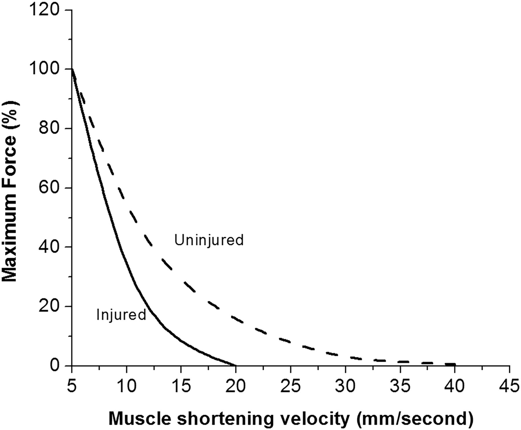
Fascicle length has a significant influence on the force–velocity and force–length relationships and, by extension, may alter muscle function. The impact of fascicle length on the force–velocity relationship has been investigated previously in the feline semitendinosus.79 This muscle has a proximal and distal head, separated by a thick tendinous inscription. Both portions have similar architectural characteristics, differing only in the length of their fascicles, with the distal head containing significantly longer fascicles (3.93±0.1 cm) than the proximal head (2.12±0.1 cm). An in vivo comparison of the maximal shortening velocities for both of these heads showed that the distal head is able to shorten approximately twice as fast (424 mm/s) as the proximal head (224 mm/s).79 As a previously strain injured muscle possesses shorter fascicles in comparison to an uninjured muscle,37 this could lead to a reduced maximal shortening velocity of the injured muscle (figures 2 and 3).

Comparison of two different muscles with identical architectural characteristics, however one contains longer fascicles (uninjured) than the other (injured). Shorter muscles fascicles have been reported in previously injured biceps femoris long head.37 Less sarcomeres in-series (shorter fascicles) will result in a slower maximal shortening velocity.

The maximal shortening velocity of a muscle is influenced by the length of the muscle fascicle. Consider that hypothetically an uninjured muscle (i) has twice the number of in-series sarcomeres that a previously injured muscle (ii) does. At any shortening velocity, the individual sarcomeres will shorten across identical distances. However, as an uninjured muscle contains more in-series sarcomeres, the entire muscle shortens over a greater distance than one with a history of injury. As velocity is the quotient of displacement and time, if these muscles shortened over the same time epoch, an uninjured muscle will possess a greater shortening velocity.
It is also hypothesised that muscle fascicle lengths have some bearing on the force–length relationship; however, evidence in humans is limited.1 ,8 ,21 It is thought that a previously injured muscle which is identical to an uninjured muscle, however with shorter fascicle lengths, will have a reduced working range as a result of fewer sarcomeres in-series.37 ,80 This may increase the amount of work being completed on the descending limb of the force–length relationship, where a reduced force-generating capacity may result in an increased potential for muscle damage.1 ,8 This concept is supported in the literature using animal models, where an increase of in-series sarcomeres in the vasti of rats and toads resulted in maximal force being produced at longer muscle lengths when compared with the vasti with fewer in-series sarcomeres.62 ,81–83 Muscle architecture plays a role in the active portion of the force–length relationship in animals models.1 ,8 ,84 It may also play a role in the generation of passive force that is produced at longer muscle lengths, yet this requires further investigation.
Impact of muscle strain injury on architecture
Limited evidence exists to characterise the effect of injury on muscle architecture. From the available literature, the isokinetic dynamometry-derived torque-joint angle relationship has been used to postulate the effects of prior hamstring strain injury on fascicle length.9 ,85–87 These studies suggest that a shift in the angle of peak torque of the knee flexors towards shorter lengths in individuals with a previously injured hamstring, is the result of a reduction in the number of in-series sarcomeres, and a decrease in the optimum length for force production.9 ,20 ,87
Evidence for shorter fascicles in individuals with a history of strain injury has recently been provided through the use of 2D ultrasound.37 Athletes who had experienced a unilateral biceps femoris long head strain injury within the preceding 18 months, had the biceps femoris long head architecture of both limbs assessed. The previously injured muscles had shorter fascicles and greater pennation angles when compared with the contralateral, uninjured biceps femoris long head.37 Owing to a lack of prospective studies, it is unclear whether these architectural changes are the cause or consequence of injury, however, their persistence long after these athletes had returned to full training and competition schedules is intriguing. It must also be acknowledged that factors such as changes in connective tissue content/fibrosis of the scar tissue88 and damage to the intramuscular nerve branches at the site of injury89 may influence these architectural differences in individuals with a history of strain injury.
Neuromuscular inhibition after strain injury has been proposed to account for fascicular shortening following a strain injury.87 ,90 The previously injured muscle has a reduced level of activation during eccentric contractions at long muscle lengths when compared to the contralateral uninjured biceps femoris long head.86 ,90 This reduced activation, as well as the avoidance of long muscle lengths during the early stages of rehabilitation, could result in structural changes (eg, reduced muscle volume, altered architecture) that would ultimately lead to adverse alterations in function.87 Despite the best efforts during rehabilitation to include heavily loaded eccentric exercises in an attempt to restore muscle structure and function to preinjured levels,91–94 the altered neural drive and difficulty in isolating the injured muscle may limit the potency of this stimulus, and thus, limit fascicle length changes.
Possessing shorter fascicles has been suggested to increase the likelihood of microscopic muscle damage as a consequence of repetitive eccentric actions (eg, high-speed running) and, when coupled with a high frequency of training sessions, may result in an accumulation of damage.87 ,95 This accumulation of eccentrically induced muscle damage would leave the muscle more vulnerable to strain injury when it encounters a potentially injurious situation, increasing the probability of reinjury.87 It is also possible that muscle fascicle length may be a primary risk factor, and explain (at least in part) why certain athletes suffer muscle strain injuries in the first place.9 ,95
It should also be noted that a number of factors are likely to influence the risk of injury and reinjury, in addition to architectural maladaptations. For example, tendon geometry is another intrinsic risk factor that has recently been proposed to have a potential role in muscle strain injuries. The width of the proximal biceps femoris tendon has been shown to exhibit high levels of variability within healthy athletes.96 Possessing a narrow proximal tendon width has been shown to increase the tissue strains within the muscle fibres adjacent to the proximal musculotendinous junction of the biceps femoris long head during active lengthening,97 and high-speed running.98 The combination of these characteristics suggest that an athlete with a narrow proximal biceps femoris long head tendon may expose the tissue surrounding this tendon to high strains and, potentially, increase the risk for injury at this site during active lengthening or high-speed running. Additionally, eccentric strength deficits and neuromuscular inhibition might themselves elevate the risk of reinjury, perhaps in conjunction with the aforementioned architectural/anatomical factors. Much work is still required in this area to confirm this hypothesis, including prospective observations to determine if shorter muscle fascicles (fewer sarcomeres in-series) increase the risk of future injury in human muscles.
Summary
Architectural characteristics of skeletal muscle characteristics can be assessed using multiple methods; of these 2D ultrasound is the most efficient and cost effective. Moreover, architecture displays plasticity in response to different stimuli, which can partly explain changes in function following training and immobilisation. Previously injured muscles have significantly shorter fascicle lengths than uninjured muscles. We present an argument as to how variations in architecture may impact function. However, no research has examined the effect that fascicle lengths have on the risk of injury. The role of architectural characteristics in muscle strain injury aetiology currently remains unknown. We recommend that investigators explore the relationship between muscle architecture and strain injury with a view to ultimately assisting in prevention of muscle strain injury and reinjury.
How might it impact on clinical practice in the future?
Injury prevention and rehabilitation strategies should consider structural adaptations.
The potency of the stimulus required to bring about structural changes is influenced by the contraction mode, velocity and muscle length. The impact of these variables will differ between muscle groups.
All of these variables must be considered when designing rehabilitation and prevention programs.
What are the findings?
Skeletal muscle architecture can be assessed using many methods including two-dimensional ultrasound, MRI and cadaveric observation.
The characteristics of muscle architecture are plastic in nature and respond to various stimuli, such as resistance, training, interventions and immobilisation.
The extent of these architectural alterations are reliant on various factors including the muscle being targeted, the range of motion/joint position during the intervention, contraction mode of training, and the velocity of the contractions.
There is only limited evidence as to how injury may alter muscle architecture and ultimately function, and conversely, the role that these characteristics may play in the aetiology of a strain injury is also unknown.
References
Footnotes
Twitter Follow Ryan Timmins at @ryan_timmins, Anthony Shield at @das_shield, Morgan Williams at @drmorgs, Christian Lorenzen at @athleticexcel and David Opar at @davidopar
Contributors RGT was primarily responsible for the determining the review design and wrote the manuscript. MDW, AJS, DAO and CL were involved in the review design and assisted in writing the manuscript.
Competing interests None declared.
Provenance and peer review Not commissioned; externally peer reviewed.