Article Text

Abstract
Objectives: To investigate the trunk strength of elite rowers and the impact of low back pain on these measures in order to determine if asymmetries or weakness were present.
Methods: Twenty two elite rowers were recruited: 13 reported previous low back pain, five current low back pain, and the remainder had no history of low back pain. All subjects were scanned during simulated rowing in an interventional open magnetic resonance imaging scanner. In each simulated rowing position, axial scans were obtained at the level of the L4–5 and L5–S1 disc interspace to determine the cross sectional area of the posterior trunk muscles.
Results: Considerable differences were observed between the three groups of rowers. In contrast with expectations and previous literature, the trunk muscles of rowers with low back pain had significantly larger cross sectional areas (p<0.001). No left/right asymmetries were observed and no differences between oarside and non-oarside in terms of muscle cross sectional area.
Conclusion: These findings suggest that low back pain in rowers does not arise as a result of muscle weakness.
- rowing
- back muscle
- cross sectional area
- multifidus
- low back pain
Statistics from Altmetric.com
Competitive rowing is a strenuous sport which requires high levels of dedication. It is an endurance sport associated with long hours of intensive training both on and off the water. Roy et al1 suggested that people with fatigue resistant back muscles and general physical fitness have fewer back problems, but the most common injury in rowers is low back pain.2–6 There is concern that the incidence of low back pain is rising, particularly in club rowers; however, further work is required to substantiate this. Whether or not it is increasing in incidence, low back pain is a considerable problem in the rowing world. It is unclear why such injuries occur, although the many speculations include poor rowing technique and weight training skills, inadequate stretching and flexibility, and changes in the equipment.6–10
In people with low back pain, the role of the paraspinal muscles with respect to stability and functional movement has been stressed.11–14 Particular emphasis has been placed on the multifidus muscle, the larger and most medial of the lumbar back muscles. Isolated unilateral wasting of this muscle has been observed in patients with low back pain.11 Since this initial work, it has been shown that recovery of this muscle is not spontaneous after remission of symptoms,15 and this may therefore be a factor in re-injury. However, the role of reduced muscle strength as a causative or predisposing factor in back disorders is controversial.16
Little is known about the strength of the back muscles in rowers; a pilot study investigating global parameters of strength did note that rowers did not have stronger backs than control subjects, although they exhibited greater strength in the thigh muscles.17 The significance of this finding with respect to back pain is not clear.
Rowing is an asymmetric activity which involves loading the back in a rotated and flexed position, factors already identified in back pain.11,16,18–22 Repetition of an asymmetric activity can lead to the development of muscle asymmetry and injury, if not addressed by appropriate training methods. Hides et al11 noted right to left differences in terms of muscle cross sectional area in subjects with and without low back pain, suggesting that muscle asymmetry may be important in the development of low back pain in the general population. Parkin et al17 noted a left/right asymmetry in muscle activity during isometric contraction of the back extensor muscles.
As well as bilateral asymmetry, imbalances can occur between the agonist and antagonist muscles. Motion studies have noted changes in the motion of the pelvis during rowing in rowers with low back pain,23 which may be caused by an imbalance of back flexors and extensors and the muscles acting at the pelvis. However, relatively few studies have investigated the relation between imbalance in muscle strength and the occurrence of injuries. Previous studies have investigated muscle weakness and imbalance after injury and surgery and suggested that imbalance is associated with injury and recurrence of injuries.24–26
We examined measures of cross sectional area of the muscles acting directly on the lumbar spine (the multifidus, erector spinae, and iliopsoas) during simulated rowing in elite oarsmen with and without low back pain.
METHODS
Study population
Twenty two elite rowers ranging from international under 23 to senior I open oarsmen were recruited primarily from the Imperial College Boat Club. The mean (SD) age of the subjects was 22.6 (4.3) years. Ten subjects rowed stroke side and 12 bow side. All had been rowing for four years or more. Thirteen subjects (mean age 23.2 (5.3) years, mean weight 87.8 (8.5) kg) reported previous low back pain which had required non-surgical intervention and had resulted in time off training, five subjects (mean age 22.0 (1.8) years, mean weight 88.7 (6.9) kg) reported current low back pain preventing full training, and four had no history of low back pain (mean age 21.0 (2.2) years, mean weight 83.4 (3.2) kg).
Imaging
Subjects were scanned using a General Electric Signa SP10 interventional magnetic resonance imaging (MRI) scanner (Milwaukee, Wisconsin, USA). This is an open configuration MRI scanner consisting of two connected but opposing ring “doughnut” magnets. The gap between these magnets is 56 cm generating a uniform field of 0.5 T. A transmit receive flexible coil was secured around the subject's waist and lumbar spine, and a multicoil magnetic resonance tracking device was positioned in line with the subject's lumbar spinous processes. Subjects were scanned with an FSPGR sequence. The parameters set were: time of repetitions (TR) 14.6; time of excitations (TE) 7.3; scan time two seconds; flip angle 60°; thickness, 10 mm; field of view (FOV) 30 cm; matrix 256 × 128; number of excitations 1. This was performed in conjunction with the magnetic resonance tracking programme (General Electric) via a Sun SPARC workstation (Sun Microsystems Corporation, Mountain View, California, USA), which permitted the subject's spine to be tracked within the scanner.27
Protocol
An MRI compatible wooden rowing jig was constructed which permitted the simulation of four key stages in the rowing stroke (the catch, early and late drive, and the finish) within the scanner.23 This study focused on simulation of the catch position within the scanner. Subjects were asked to adopt their usual position at the catch phase of the stroke, the length of the oar was adjusted accordingly, and they were asked to pull on the oar (thus loading the spine and contracting the muscles) as they would while rowing. With the rower in this position, a sagittal scan of the lumbar spine was performed to localise the region of interest followed by a series of axial scans through the intervertebral junction of the L4–5 disc interspace and the L5–S1 disc interspace. Subjects were asked to remain as still as possible during scanning.
Image analysis
Images were analysed on a conventional workstation to allow the measurement of muscle cross sectional area. Previous studies have shown a good correlation between MRI measures of cross sectional area and anatomical measurement.28,29 Muscles assessed were the multifidus, erector spine muscle group at the L4–5 and L5–S1 level, and the iliopsoas at the L4–5 level.
Statistical analysis
The statistical analysis was performed using the statistical package Stata, version 6 (Stata Corporation, College Station, Texas, USA) on a personal computer. A two way analysis of variance was used to investigate if any differences existed between the three population groups. A series of covariates were considered in the analysis of variance in terms of their influence on cross sectional area, including effects of age, stroke side, and side of the body. The statistical threshold was set at p<0.05. Orthogonal contrasts and multiple regression analysis were then used to locate where any differences noted by the analysis of variance lay.
RESULTS
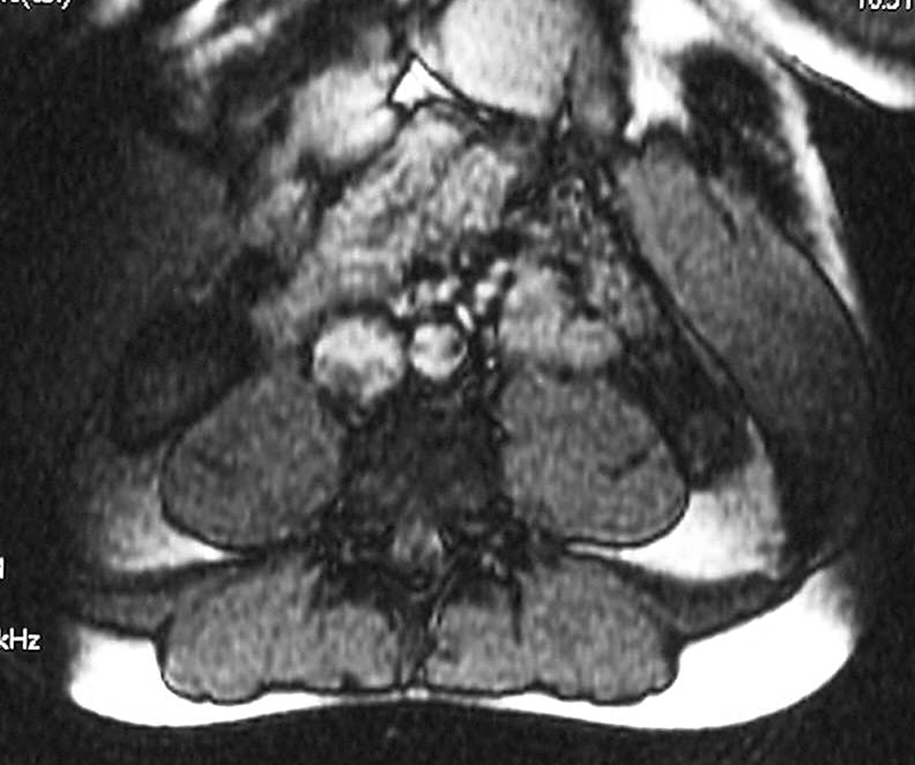
The imaging protocol resulted in clear images of the lumbar spine muscles, including the erector spinae, multifidus, and iliopsoas (fig 1), from which measures of cross sectional area could be obtained.

Axial view of the lumbar muscles at the L4–5 intervertebral junction during a contracted magnetic resonance imaging scan.
Table 1 summarises the measurements of cross sectional area obtained on both the left and right sides of the spine.
Measures of cross sectional area (mm2) in each of the muscle groups considered
Statistical analysis revealed significant differences with respect to cross sectional area between the three study populations. In terms of the multifidus muscle, rowers with back pain (both current and previous) were noted to have significantly larger muscles than those without back pain, this being most prominent in those with a previous history of low back pain (p<0.0001) (fig 2). Significant differences were also observed between subjects with a current history and those with a previous history of low back pain (p<0.001).
Cross sectional area (mm2) of the multifidus muscle in rowers with no, current, or previous low back pain.
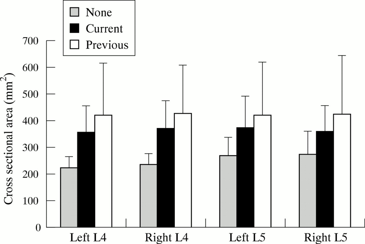
As with the multifidus muscle, significant differences were observed between the three groups when the cross sectional area of the erector spinae muscle was considered. At the L4–5 level, subjects with low back pain (both current and previous) had significantly larger muscles than those with no history of low back pain (p<0.001) (fig 3). No differences were observed between subjects with current and those with previous low back pain. However, at the L5–S1 level, these differences were reversed, with a slight tendency for those with no history of low back pain to have larger muscles. This trend was significant when subjects with no history of low back pain were compared with those with a previous history of low back pain (p<0.05).

Cross sectional area (mm2) of the erector spinae muscle group in rowers with no, current, or previous low back pain.
When the iliopsoas muscle was considered, a similar trend was observed, with subjects with a history of low back pain (both current and previous) having larger cross sectional areas (p<0.0001). However, no differences were observed between the two back pain groups. When the ratio of total back extensor muscle—that is, erector spinae and multifidus—to iliopsoas cross sectional area was compared, subjects with a previous history of low back pain had a significantly lower ratio than those with no history of low back pain (p<0.05).
Muscle cross sectional area was influenced by age, with a decrease in muscle size with an increase in age. In some rowers, there appeared to be left and right asymmetries. However, this was not the global trend, and no significant left/right asymmetries were observed for any of the muscle groups. Similarly there were no significant asymmetries observed when oarside and non-oarside were compared.
DISCUSSION
Controversy exists about the strength of the trunk muscles and their association with both the presence and incidence of low back pain. Some studies show little or no association between strength and low back pain,19,20 whereas others show notable weakness of the trunk muscles.1,16,21,22 Biering-Sorensen et al30 reported that weak trunk muscle is one of the greatest risk indicators for a first experience of low back pain, although more recent studies have disagreed with this.31 Despite this controversy, exercises to strengthen trunk muscles are commonly recommended not only as a treatment for low back pain, but as a possible preventive measure.32
Little is known about the capabilities of the trunk muscles of athletes, and the role of these muscles in the generation of low back pain. Many advocate that the treatment of athletes with low back pain should include exercises to correct imbalances and weakness.33 Muller et al34 noted that elite rowers had greater isometric torque of the trunk muscles than both control subjects and other sportspeople such as swimmers and tennis players. However, this is in contrast with the finding of Parkin et al,17 who noted no differences between elite rowing and control populations. Investigations of strength in the rowing population have not been performed.
This study has investigated measures of cross sectional area of the back muscles in a group of elite rowers with and without low back pain. Measures of cross sectional area have often been used to determine the force generating capacity of a muscle group.35 Comparisons of these three groups of rowers produced some unexpected results and suggested that rowers with back pain have larger back muscles, and thus greater strength, than rowers without low back pain. This is in contrast with the findings for the general low back pain population. It is not known if this observation is a cause or an effect of low back pain. In addition, the comparatively small study population may have influenced the findings of the study. The increased strength in the spinal muscles may be a consequence of poor technique, the rowers with back pain predominantly using their backs to generate force during the stroke rather than their legs. Previous studies have suggested that there may be an alteration in the lumbopelvic rhythm of rowers with back pain that may lead to hypertrophy of the back muscles.19 It has also been proposed that patients with low back pain present with deficits in motor control as opposed to impairments in strength36,37; if so, these changes in strength may be irrelevant. Other authors have speculated that it is fatigue not strength of the back muscles that is important.4 This was not addressed in this study.
Take home message
Low back pain in elite oarsmen does not appear to be the result of weakness or asymmetry of the multifidus or erector spinae muscle group.
Muller et al34 noted that the extension/flexion ratio of the trunk was lower in subjects with higher rowing performance, suggesting high levels of trunk flexor strength. The influence of injury on this ratio was not assessed, because the study was unable to assess the abdominal mechanism. Consequently changes in the strength of the transversus abdominus, an important spinal stabilising muscle, have not been examined. Assessment of the cross sectional area of this muscle may provide further information to account for the unexpected finding of increased strength in the posterior stabilising muscles of the rowers with low back pain.
The asymmetric nature of rowing has been speculated to lead to the development of muscle asymmetries in the spine. Parkin et al17 investigated muscle activity patterns during isometric trunk extension and noted left and right asymmetries in the rowers. However, we were unable to detect any differences in cross sectional area between the left and right sides. Similarly no differences were detected when oarside and non-oarside muscles were compared.