Article Text

Abstract
Background: Overuse injury to the patellar tendon (patellar tendinopathy) is a major reason for interrupted training and competition for elite athletes. In both sexes, the prevalence of unilateral and bilateral tendinopathy has been shown to differ. It has been proposed that bilateral pathology may have a different aetiology from unilateral pathology. Investigation of risk factors that may be unique to unilateral and bilateral patellar tendinopathy in female athletes may reveal insights into the aetiology of this condition.
Objectives: To examine whether anthropometry, body composition, or muscle strength distinguished elite female basketball players with unilateral or bilateral patellar tendinopathy.
Methods: Body composition, anthropometry, and muscle strength were compared in elite female basketball players with unilateral (n = 8), bilateral (n = 7), or no (n = 24) patellar tendinopathy. Body composition was analysed using a dual energy x ray absorptiometer. Anthropometric measures were assessed using standard techniques. Knee extensor strength was measured at 180°/s using an isokinetic dynamometer. z scores were calculated for the unilateral and bilateral groups (using the no tendinopathy group as controls). z scores were tested against zero.
Results: The tibia length to stature ratio was approximately 1.3 (1.3) SDs above zero in both the affected and non-affected legs in the unilateral group (p<0.05). The waist to hip ratio was 0.66 (0.78) SD above zero in the unilateral group (p<0.05). In the unilateral group, leg lean to total lean ratio was 0.42 (0.55) SD above zero (p<0.07), the trunk lean to total lean ratio was 0.63 (0.68) SD below zero (p<0.05), and leg fat relative to total fat was 0.47 (0.65) SD below zero (p<0.09). In the unilateral group, the leg with pathology was 0.78 (1.03) SD weaker during eccentric contractions (p<0.07).
Conclusions: Unilateral patellar tendinopathy has identifiable risk factors whereas bilateral patellar tendinopathy may not. This suggests that the aetiology of these conditions may be different. However, interpretation must respect the limitation of small subject numbers.
- patellar tendinopathy
- anthropometry
- body composition
- female athletes
- basketball
Statistics from Altmetric.com
Overuse injury to the patellar tendon (patellar tendinopathy) is common among athletes (particularly at elite levels) and is a major reason for interrupted training and competition.1,2 It is notoriously difficult to treat and may result in untimely retirement from competitive sport.3 Despite being first described in 1973,4 little is known about the aetiology of this condition. Strength and anthropometric measures have been associated with symptomatic patellar tendinopathy in male athletes, although the findings are equivocal.5–11 Similar data in female athletes are scarce.
The prevalence of unilateral and bilateral patellar tendinopathy has been shown to differ between the sexes; bilateral tendinopathy is equally prevalent, but unilateral tendinopathy is twice as common in males as females.12 This suggests that bilateral pathology may have a different aetiology from unilateral pathology. This is supported by evidence that female athletes with unilateral patellar tendinopathy had significantly less posterior leg flexibility than subjects with bilateral patellar tendinopathy and subjects with normal tendons.5
The reason for the different prevalence and presentation of unilateral and bilateral tendinopathy may be individual and inherent characteristics. The genetic profile is associated with severe collagen disease,13 and, although yet to be shown, collagen changes in some presentations of tendon disease may also have a genetic link. In the Achilles tendon, the ABO blood grouping has been associated with pathology; subjects with bilateral tendon rupture had the highest frequency of type O blood (71%) followed by unilateral tendon involvement (53%); both were higher than the general population (31%).14
Investigation of risk factors that may be unique to unilateral and bilateral patellar tendinopathy in women may reveal insights into the aetiology of tendon pathology in these two groups. Thus we asked the following question: are elite female basketball players with unilateral or bilateral patellar tendinopathy distinguished by differences in anthropometry, body composition, or muscle strength?
METHODS
Two Women’s National Basketball League (WNBL) and six Australian Basketball Association (ABA) clubs were invited to participate in this research. Subjects were considered suitable for participation if they were currently engaged in full training and match responsibilities, had not had knee surgery in the preceding six months, and had never had surgery on the patellar tendon. Current symptoms of patellar tendinopathy did not exclude subjects if they met the above criteria. The faculty human ethics committees at La Trobe University, Deakin University, and the Radiation Safety Unit approved all procedures. All subjects gave written informed consent.
Subjects recorded symptoms (VISA score15) and details on training, injuries, surgery, contraceptive use, and menstrual history. Physical performance, body composition, and anthropometric values were then tested. Subsequently an ultrasound examination of both patellar tendons was performed by an experienced musculoskeletal radiologist.
Physical and anthropometric testing
All physical and anthropometric testing was conducted in one session at the human movement laboratories at Deakin University by the same investigator (JEG) and a trained anthropometrist (SA). Standing and sitting heights were measured using a Holtain wall stadiometer and sitting height table (British Indicators Ltd, Burgess Hill, West Sussex, UK). Weight was measured using a Seca electronic scale (Seca Ltd, Birmingham, UK). Segmental limb lengths were measured using a Harpenden anthropometer (British Indicators Ltd); coefficient of variability was 0.1±0.1%.16
Physical performance testing measured sit and reach, vertical jump, and isokinetic quadriceps strength. Before a standard warm up and stretch, sit and reach was determined using standard procedures. Right leg, left leg, and bilateral vertical jump were then tested using a Vertec (Sports Imports Inc, Columbus, Ohio, USA). The protocol allowed no run up or arm swing during take off. A countermovement to 90° of knee flexion was allowed for all jumps. Lastly, isokinetic strength of the quadriceps was tested using a Cybex dynamometer (Lumex Inc, Ronkonkoma, New York, USA). Five familiarisation trials followed by three recorded trials were performed at 180°/s. Output was recorded as average work over the three trials.
Body composition
Body composition was analysed using a dual energy x ray absorptiometer (DPX-L; Lunar Radiation Corporation, Madison, Wisconsin, USA). Total and regional fat and lean body mass were determined using a total body scan.
Ultrasound examination
Each subject had an ultrasound examination of both patellar tendons. The examinations were performed by an experienced radiologist with additional musculoskeletal training. For the detection of hypoechoic lesions, both the intertester (κ = 1)17 and test-retest reliability (40/40)18 has been documented as excellent for a radiologist with musculoskeletal training.
Both transverse and longitudinal scans were conducted using a high resolution linear array with a 10 or 12 MHz transducer. Tendons were designated pathological if a hypoechoic lesion was visible in both the longitudinal and transverse scans. The ultrasound results for both tendons were used to assign subjects to one of three naturally occurring groups: no tendinopathy (control), unilateral tendinopathy, or bilateral tendinopathy.
Data analysis
Data for these three groups were then entered into SPSS (SPSS Inc, Chicago, Illinois, USA). Analysis was conducted using the median score of three trials for anthropometric data16 and mean of three trials for all other data. Data for the unilateral and bilateral tendinopathy groups were then expressed as z scores using the mean and standard deviation of the corresponding score from the control group. z scores for each leg were calculated from the comparable leg—that is, left or right leg—in the controls. A one sample t test was used to determine if any z scores deviated from zero. Results are reported to be significant at p<0.05, and trends are reported where p<0.10, as this allows the reader to determine both the clinical and further research significance of these findings.19 For clarity, the unilateral and bilateral patellar tendinopathy groups will be referred to as the “unilateral” and “bilateral” groups respectively.
RESULTS
Thirty nine subjects were recruited, 16 from the WNBL and 23 from the ABA. The prevalence of tendinopathy (unilateral and bilateral) did not differ between the two levels of competition. Ultrasound examination showed a unilateral hypoechoic lesion in eight athletes, bilateral lesions in seven athletes, and no lesion in 24 athletes. Subjects with one or two hypoechoic lesions did not differ from controls in age, menstrual history, height, or weight (table 1). Subjects with one or two hypoechoic lesions, however, had trained a mean (SD) of 2.6 (1.4) more hours a week than controls in the preceding one to six months and had lower VISA scores (12.1 (5.7)) than controls (both p<0.05) (table 2).
Age, menstrual history, height, weight, anthropometric data, body composition data, and strength data of female basketball players with unilateral or bilateral patellar tendinopathy and controls
Training history and VISA score for female basketball players with unilateral or bilateral patellar tendinopathy and controls
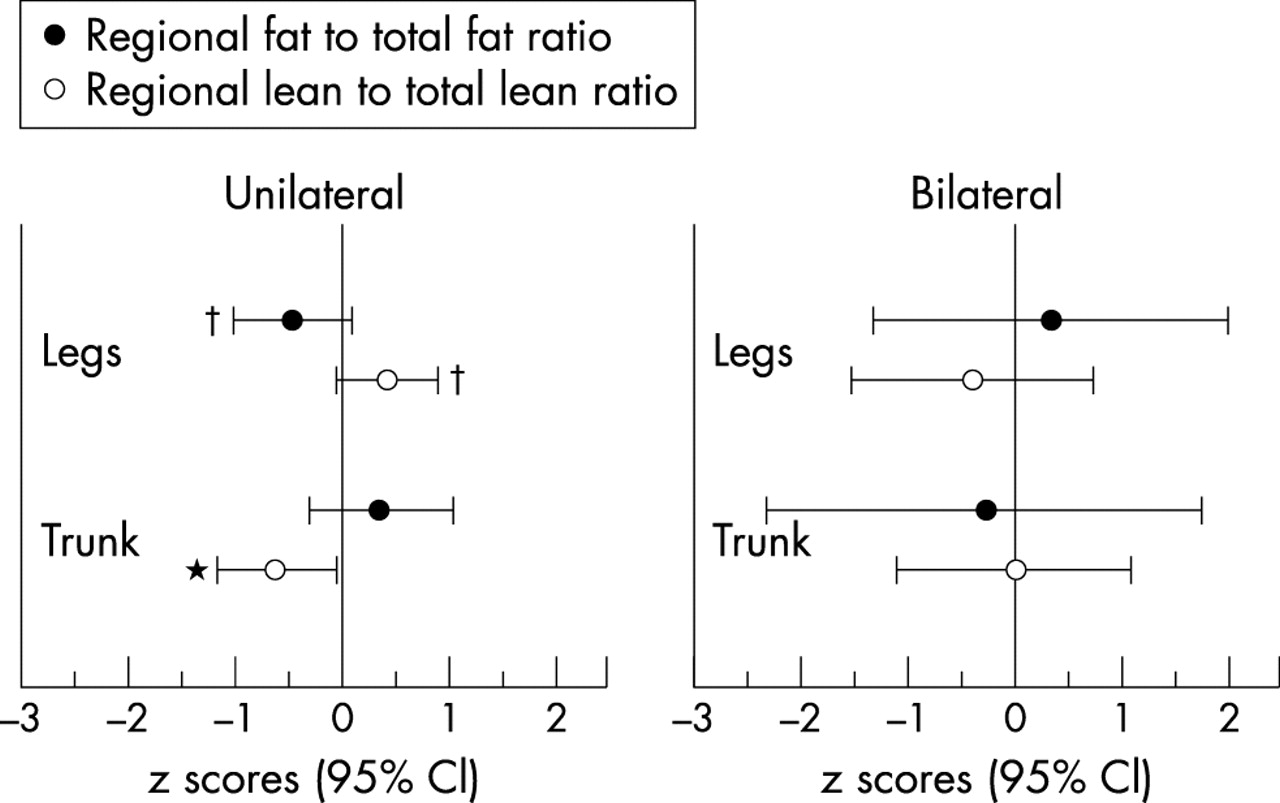
There were no detectable differences between the bilateral group and controls in any of the measured variables. In contrast, subjects in the unilateral group were distinguished from the control group by differences in anthropometry, body composition, and strength. The tibia length to stature ratio was approximately 1.3 (1.3) SDs above zero in both the affected and non-affected legs in the unilateral group (p<0.05) (fig 1). Although no differences in waist or hip girths were detected between the groups, the waist to hip ratio was 0.66 (0.78) SD above zero in the unilateral group (p<0.05) (fig 2). The unilateral group leg lean to total lean ratio was 0.42 (0.55) SD above zero (p<0.07). In contrast, trunk lean to total lean ratio was 0.63 (0.68) SD below zero (p<0.05) (fig 3). The reverse was evident for fat mass: the leg fat relative to total fat was 0.47 (0.65) SD below zero (p<0.09) (fig 3). In the unilateral group the leg with pathology was 0.78 (1.03) SD weaker during eccentric contractions (p<0.07).

Femur to stature ratio and tibia to stature ratio z scores for the affected and unaffected leg in subjects with unilateral and bilateral patellar tendinopathy (mean and 95% confidence interval (CI)). *Significantly different from zero, p<0.05.
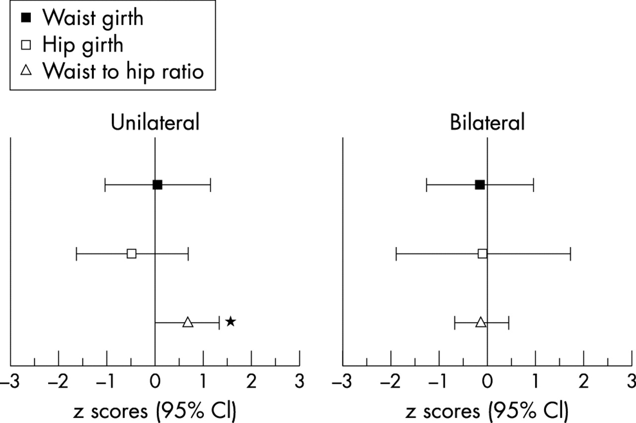
Waist girth, hip girth, and waist to hip ratio z scores in subjects with unilateral and bilateral patellar tendinopathy (mean and 95% confidence interval (CI)). *Significantly different from zero, p<0.05.
Regional fat to total fat ratio and regional lean to total lean ratio z scores for the legs and trunk of subjects with unilateral and bilateral patellar tendinopathy (mean and 95% confidence interval (CI)). *Significantly different from zero, p<0.05; †Significantly different from zero, p<0.10.
DISCUSSION
In this study of elite female basketball players, several anthropometric and body composition variables distinguished athletes with one hypoechoic tendon from controls. No measured variable distinguished athletes with two hypoechoic tendons from controls.
Subjects with unilateral patellar tendinopathy had a higher waist to hip ratio than controls. This indicates a larger abdominal fat distribution relative to gluteofemoral fat deposits.16 Human fat distribution is controlled by a complex interaction of hormones and is particularly influenced by the female sex hormones (oestrogen and progesterone), but the mechanisms whereby hormones control this fat distribution are unclear.20
Hormone concentrations may also influence susceptibility to tendinopathy. Oestrogen has been reported to directly affect fibroblast proliferation and collagen synthesis,21–24 and menopause is associated with an increase in Achilles tendon ruptures, a finding that is not paralleled in men of an equivalent age.25 This study did not examine hormone concentrations and thus this hypothesis cannot be substantiated. However, these data together support the notion that hormone concentrations that may affect connective tissue are characterised by a redistribution of body fat to the truncal region. Furthermore, combining the decrease in trunk lean to total lean ratio along with trends for leg lean and fat observed within the data suggest that subjects with unilateral tendinopathy have unique body compositions. Together these data indicate an android distribution of both fat and lean mass in subjects with unilateral patellar tendinopathy.
Patellar tendinopathy appears to result from multiple risk factors. Often the presence of pathology has attributed been to differences in limb lengths leading to poor movement mechanics or biomechanical factors.26,27 We found that the players with unilateral patellar tendinopathy had a longer tibia length relative to stature. This has not previously been reported, but there is evidence that an increase in both tibial and femoral lengths are associated with overuse knee injuries.28
The simplest explanation for longer tibiae in subjects with unilateral patellar tendinopathy is that altered lower limb biomechanics increases the stress on the patellar tendon and leads to tendinopathy. This appears unlikely, however, as only one tendon was affected while both tibiae were longer than the control group. Furthermore, all subjects with unilateral tendinopathy played right handed while equal numbers of left and right tendons were affected. Tibial length and unilateral patellar tendinopathy may be parallel phenomena both arising from an underlying characteristic in the individual. The relation appears complex, as the increase in length is specific to the tibia in relation to stature. To affect the length of long bones and influence tendon collagen and/or matrix morphology suggests a fundamental characteristic, which may include individual genetic or hormonal profile. This finding, however, remains unclear.
The knee extensor mechanism of the affected leg in the unilateral tendinopathy group showed a trend toward decreased eccentric strength at 180°/s. Three observations infer that this finding results from inhibition of eccentric contractions rather than muscular atrophy. Firstly, these subjects have more leg musculature than controls. Secondly, knee extensor strength does not differ from controls for concentric contractions. Thirdly, elite basketball players do not tend to develop one sided weakness as the ability to lead with or jump from either leg is a highly valued attribute. Decreased eccentric strength may initially protect the tendon from abusive stress levels (highest during eccentric contractions) but a decrease in controlled muscle strength may ultimately be detrimental to tendons.29
Limitations of the study
A number of the topics in the discussion led to the suggestion that female sex hormones may influence the risk of pathology in these athletes. This notion is speculative as sex steroids were not measured in this study and this warrants further investigation. Furthermore, the study design was cross sectional with relatively small subject numbers. Notwithstanding this, a number of significant results were reported. These data, however, need to be confirmed using large sample sizes.
CONCLUSION
These data suggest that unilateral and bilateral patellar tendinopathy have unique aetiology. Several variables in the group with unilateral patellar tendinopathy differed significantly from controls, whereas no variable distinguished subjects with bilateral patellar tendinopathy from controls. In fact, those variables that reached significance in the unilateral group tended toward the opposite direction in the bilateral group. Although further research is required to validate these findings in larger samples, across sports, and in male subjects, these data suggest that unilateral and bilateral patellar tendinopathy warrant separate investigation.
Acknowledgments
We thank the players and coaches of the Bullen Boomers (WNBL), Dandenong Rangers (WNBL and ABA), Eltham Wildcats (ABA), Hume City Broncos (ABA), Kilsyth Lady Cobras (ABA), Knox Raiders (ABA), and Nunawading Spectres (ABA).