Article Text

Statistics from Altmetric.com
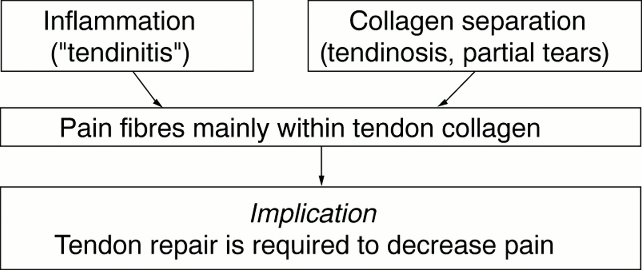
Traditional dogma would have it that pain in tendinopathy arises through one of two mechanisms. Firstly, it may result from inflammation in “tendinitis”. Secondly, it may be due to separation of collagen fibres in more severe forms of tendinopathy. The latter situation parallels the mechanism of pain with collagen separation after an acute grade I or II ligament injury (fig 1).
Despite the wide acceptance of these two classical models of pain production, a number of studies provide data inconsistent with either theory. Consider first the inflammation mechanism. Histopathological examination of surgical specimens from patients with chronic tendon pain are devoid of inflammatory cells.1 This applies to tissue from the Achilles, patellar, lateral elbow, medial elbow, and rotator cuff tendons. Furthermore, prostaglandin E2 (a marker of the inflammatory process) is no more abundant in patients with Achilles tendon pain than in normal controls.2
Unfortunately, the collagen separation theory does not hold up under scrutiny either. The following five observations about pain and collagen in the patellar tendon are inexplicable. (a) Patients who have patellar tendon allograft anterior cruciate ligament reconstruction have minimal donor site knee pain, yet collagen has been excised. (b) Such patients are generally pain-free (and back at sport) despite the persistence of abnormal collagen for two or more years.3,4 (c) Similarly, after open surgery for jumper's knee, the imaging appearance of the tendon—that is, collagen status—does not correlate consistently with knee pain.5 (d) Patients with jumper's knee can also be treated by an arthroscopic debridement of the infrapatellar fat pad and the posterior border of the patellar tendon without operation on the collagen defect in the tendon itself.6 (e) Large asymptomatic ultrasonographic hypoechoic regions (abnormal collagen) can be found in patellar tendons of some athletes who have never had a history of jumper's knee.7,8
Such discrepancy between collagen structure and pain is not confined to the patellar tendon. Patients with partial (non-perforated) rotator cuff tears were found to have more pain than those with complete perforations9 despite the former having less collagen damage. Clearly there is more to tendon pain than discontinuity of collagen per se.
Nociceptors provide significant afferent pain pathways. In the knee, they are located in the retinaculum, fat pad, synovium, and periosteum,10 and all these structures may play a role in the tendon pain pathway. Biochemical irritants may include extravasation of glycosamines, especially chondroitin sulphate,11,12 from damaged tendon.
The five observations listed above can be explained with what we term a “biochemical” hypothesis (fig 2). We speculate that the pain of patellar tendinopathy is largely due to biochemical agents irritating nociceptors located in the fat pad immediately posterior to the patellar tendon. In 39 cadaver dissections of the proximal patellar tendon,13 we consistently identified a thin layer of fat adherent to the posterior portion of the patellar tendon. In the corresponding tissue specimens from patients operated on for chronic jumper's knee, this fat tissue contained increased Alcian blue stain (and thus glycosaminoglycans), presumably leaked from the adjacent region of tendinosis.
To our knowledge, the key irritant biochemical agent has not yet been identified, and this presents a challenge for tendon biochemists. Using microdialysis, Alfredson recently identified an abnormal amount of the excitatory neurotransmitter, glutamate, in subjects with painful Achilles tendinopathy.2 Until these histopathological and biochemical findings are correlated with some measure of pain, we can only speculate as to whether they are causative, or merely byproducts of nearby tendinosis.
Of interest, in the rotator cuff pain and pathology study quoted above,9 collagen damage was inversely related to pain, but the presence of substance P (a nociceptive neurotransmitter) was significantly associated with pain. Nerve fibres immunoreactive to substance P were localised around vessels in the subacromial bursa and in the non-perforated rotator cuff.9
Although the data presented may suggest a biochemical cause of pain, other workers consider mechanical impingement of the fat pad as a cause of anterior knee pain. The Australian physiotherapist, Jenny McConnell, recognised fat pad impingement as a cause of anterior knee pain (not necessarily tendon pain) over 10 years ago. Johnson proposed that impingement caused the pain of patellar tendinopathy.14 The infrapatellar fat pad is an extremely sensitive region15 and contains a large number of nociceptors, but as tendon pain occurs at many anatomical sites, it does not appear logical that a structure related to only one tendon—that is, the patellar fat pad—would necessarily play a unique role in a problem as widespread as tendinopathy. Further, the clinical observation that the pain of jumper's knee does not disappear and may actually increase when palpation is performed with the knee in full extension would appear to argue more for a biochemical than a mechanical cause of pain in tendinopathy. Nevertheless, the jury requires more evidence.
If our biochemical hypothesis proves to have some validity, it would have significant clinical and research implications. In clinical management, the aim of treatment would be to modify the biochemical milieu, rather than to focus on reducing inflammation or necessarily augmenting collagen repair. Collagen repair may, of course, improve the biochemical milieu and thus explain why eccentric strengthening programmes can help.16 Researchers would be encouraged to pursue a pharmaceutical approach focused on reducing the irritant (but not necessarily inflammatory) biochemical compounds around the tendon. Surgery may play a role through denervation. Thus, if sports medicine researchers collaborate with basic scientists who understand pain physiology, knowledge will be advanced in both fields, and we will progress toward the goal of alleviating the pain of what is often structurally rather a trivial problem.

The classical “inflammatory” and “structural” tendon pain models.

Contemporary “biochemical” tendon pain model.
References
Linked Articles
- Correction