Article Text

Statistics from Altmetric.com
Perhaps the hallmark study in human exercise physiology was performed by Nobel Laureate Professor AV Hill on himself in Manchester, England in the early 1920s. Hill circled an 88 metre grass running track at three different speeds each for 4 minutes while he measured his average oxygen consumption every 30 seconds (Hill and Lupton;1 fig 2 of that paper). He concluded that his oxygen consumption reached a maximum at 16 km/hour “beyond which no bodily effort can drive it”.2 (page 1661) This experiment established the single most popular test in the exercise sciences – the progressive exercise test for the measurement of the maximum oxygen consumption (VO2max). The experimental protocol in this test forces the subject progressively to increase the work rate until voluntary exhaustion.
According to the modern interpretation,3–14 the outcome of this test defines the limits of the human cardiorespiratory system, because it apparently terminates when the cardiac output reaches a maximum value.9 11 13 It also established a model to explain the biology of human exercise performance, for if the cardiovascular system determines maximal exercise performance, then it must also determine performance during many other forms of exercise, as suggested by Bassett and Howley9 and others.15 16 Thus, champion athletes able to run very fast for long distances do so because of the metabolic consequences in their skeletal muscles of their superior cardiovascular function,9 15 16 even though they exercise at intensities below that at which the VO2max is reached and at which, according to that model, cardiovascular function is not maximal and cannot therefore, by definition, be the “limiting” factor.
This explanation seems paradoxical because, if correct, it predicts that athletes should be able to exercise more vigorously and for longer simply by increasing their (submaximal) cardiac outputs until maximal values are reached. Only then should exhaustion occur. Alternatively, any athlete chasing the race leader should be able to increase the cardiac output to a maximal value and so pass the leading athlete, who is exercising at a lower and submaximal cardiac output. However, this does not happen; as is well known, prolonged exercise always terminates at submaximal levels of cardiac output.
It is my opinion that the manner in which the VO2max test is conducted has encouraged a reductionist mindset, which teaches that the “limits” of exercise performance can be explained by one or two cardiovascular variables, such as the cardiac output and muscle blood flow,15 but the VO2max test includes three components that are foreign to all forms of freely chosen exercise.
First, the tested subject does not know the expected duration of the exercise bout when it begins. Accurate knowledge of the exercise duration optimises the exercise performance.17 Second, the intensity of the exercise increases progressively, sometimes rapidly8 from low to “maximal” work rates. Humans do not usually exercise this way. Third, the tested subject cannot regulate the exercise intensity except by choosing when to stop. This adds a subjective component to the test, as the athlete’s conscious brain makes the final decision when to terminate the exercise. Thus psychological and not purely physiological factors can presumably influence that decision.18
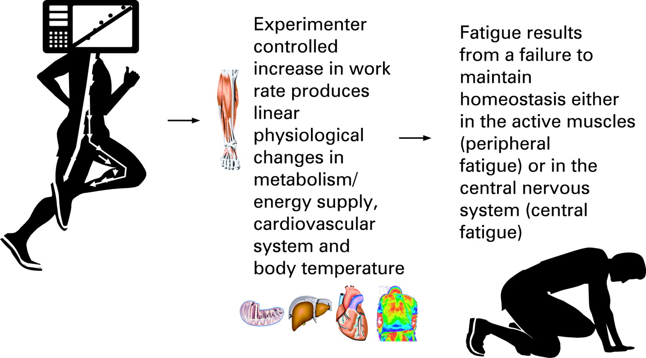
More to the point, a characteristic of freely chosen exercise is the choice of different pacing strategies that change continuously from moment to moment.19–21 Unique, constantly changing pacing strategies are most likely produced by a central motor command that continually modulates the number of motor units recruited in the exercising limbs. However, during the VO2max test, this critical brain function cannot be evaluated, because the change in work rate is preset and immutable, thereby controlling the tested subject’s level of central motor output in an unnatural way (fig 1).
Finally, the VO2max test has produced an unusual definition of the intensity at which exercise is performed, because the intensity is expressed relative to that at which the VO2max occurs. Workloads beyond those reached during the VO2max test are defined as “supramaximal”;5 12 however, this does not make sense, because a (lower) exercise intensity cannot be maximal if a higher exercise intensity can be achieved, even if under different circumstances.
Experimental models such as the VO2max test have their uses, because they can define the maximal capacity of each human for oxygen use,15 but their generalisability must be carefully scrutinised. Thus one must ask: is it appropriate to explain the physiological factors determining human exercise performance according to an experimental model of exercise in which (1) humans do not usually engage and (2) the brain of the tested subject does not set the pacing strategy as is usual in freely chosen exercise? If we base our interpretations exclusively on a testing model that is so unnatural as to exclude the usual function of the brain during exercise, we may miss the obvious. Thus, this traditional reductionist explanation of the factors limiting the VO2max excludes any possible contribution of the brain and central motor command.3–9 11–14 The point is, as Kayser18 reminds us, that exercise begins and ends in the brain. Thus, before any movement can occur, the appropriate number of motor units in the exercising skeletal muscles must first be activated by the central nervous system. As a result, the power output of the exercising limbs increases, raising the whole body oxygen consumption consequent to metabolite-induced arteriolar vasodilation, which directs the increase in blood flow to the exercising muscles. Thus, as is usually taught in standard textbooks of human physiology22 increases in blood flow and cardiac output during exercise are the consequence and not the cause of the increase in power output by the exercising muscles. Attempts to point this out are usually dismissed out of hand.23
However, the logical point is that this critical role of central motor command cannot be identified if its most important function — the setting of the pacing strategy — is the controlled variable in the experimental model — the VO2max test — used to predict athletic ability and to determine the factors that limit human exercise performance. Rather as the subject’s pacing strategy is predetermined during the VO2max test, the popular use of this specific test must entrench the conclusion that exercise performance is determined by the cardiovascular system, as it is the only obvious candidate other than central motor command.
However, the absence of proof for an effect does not prove the absence of that effect. If central motor command plays no part in determining the VO2max, then the VO2max test must terminate because a severe degree of peripheral fatigue has developed in the exercising muscles as recently acknowledged: “athletes stop exercising at VO2max because of severe functional alterations at the local muscle level due to what is ultimately a limitation in convective oxygen transport, which activates muscle afferents leading to cessation of a central motor drive and voluntary effort”.15
This novel explanation differs from the more usual theory that maximal exercise performance terminates purely as a result of the development of “peripheral fatigue”; however, it is closer to Hill’s original theory that maximal exercise performance is limited by a “governor” in either the brain or heart,24 the function of which is to terminate exercise before severe myocardial ischemia causes irreversible heart damage or even death.
Remarkably, this presumption that the VO2max test produces “severe” alterations in skeletal muscle function has only just been tested. Thus Thomas and Stephane25 have reported that the function of the quadriceps femoris muscle was little affected by the performance of a VO2max test of ∼13 minutes that terminated at a mean power output of 381 W and a mean VO2max of 75 ml/kg/min. They also showed that oxygenation of the pre-frontal cortex fell during exercise. They propose that a decrease in oxygenation of the pre-frontal cortex may modify central motor output to the exercising limbs, causing the termination of exercise before the onset of peripheral skeletal muscle fatigue.
This interpretation differs in two important ways from the new model of Levine,15 which theorises that (1) sensory feedback from fatigued skeletal muscles, not from an increasingly hypoxic prefrontal cortex, causes a failure of central motor output and (2) that this occurs only after the peripheral fatigue has already developed. Interestingly, both models accept that the brain ultimately determines when the exercise will terminate, a significant conceptual advance.18 26 27
In a somewhat related study, Kabitz et al28 have reported that electrically stimulated diaphragmatic force output increased progressively during exercise but fell immediately exercise terminated, but only when whole-body exercise was performed.29 The authors concluded that diaphragmatic fatigue develops after, not during exercise, so that “the conventional understanding of fatigue might be flawed because it does not distinguish between the sensation itself and the physical expression of that sensation26”. Interestingly, there are studies showing that skeletal muscle contractile function may also increase during exercise,30 indicating the absence of fatigue. However, the relevance of these studies is not often acknowledged as they produce a result that is so unexpected.31
Although the mechanisms explaining the unexpected findings of Kabitz et al28 29 need to be determined, those studies should encourage exercise physiologists to re-evaluate the implications of studies showing that skeletal muscle contractile function does not necessarily fall during exercise but may sometimes increase.
Importantly, the studies of Kabitz et al28 29 raise the possibility that the use of electrical stimulation to detect the presence of “peripheral fatigue” in skeletal muscle might theoretically produce contrasting results if tested during exercise rather than after the termination of exercise. Indeed, it is clear that electrical stimulation overestimates the degree of “peripheral fatigue” that may be present at the end of exercise. Thus, for example, the study of Amann et al32 found that cyclists produced an “end spurt” in 5 km cycling time trials in which their power outputs reached higher values in the final 500 m than at any time during the trials. Furthermore, power output during the end spurt increased more than did lower limb EMG activities. This suggests an increase in power production for each unit of EMG activity—that is, an increase in the capacity of the voluntarily activated skeletal muscle motor units to produce force, hence resulting in an increased, not a reduced mechanical function. Yet, when studied 4 minutes after the end of exercise, the ability of these muscles to produce a maximal voluntary contraction had fallen by ∼10% compared with pre-exercise values. Similarly, muscle-stimulation techniques consistently found a reduction of ∼35% in skeletal muscle force production. The authors interpreted this finding as evidence that the previously exercised skeletal muscles showed a 35% increase in “peripheral fatigue”. All these discrepant findings raise the question of how the extent of “peripheral fatigue” should be quantified during or after whole body exercise.
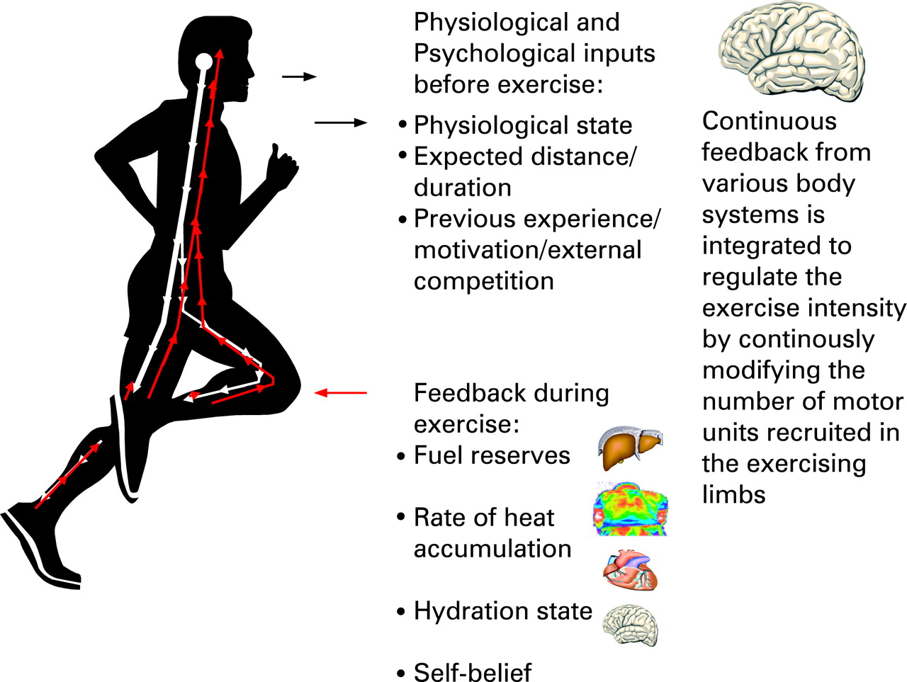
More to the point, if it is correct that cardiovascular function determines maximal exercise performance when the work load is set by the experimenter,15 then the cardiovascular system must also explain the performance of humans when they set their own pacing strategies19–21 (fig 2). Such strategies are present essentially from the first muscular contraction33 and are constantly changing.20 21 There is also usually an “end spurt” shortly before the end of exercise.32–34 As the pacing strategy is evident from the onset of exercise, it must be anticipatory and part of a feed-forward control mechanism (fig 2).19 21 35 36

How can the cardiovascular system determine the exercise performance “in anticipation” even from the very onset of exercise? How does it sanction moment-to-moment changes in power output?20 21 And how does it produce the “end spurt”,36 given that neither the heart nor the exercising limbs can ever know when the exercise will end?
More logically, central motor command determines the exercise intensity and sets the pacing strategy at the start of exercise, based largely on the expected duration of the exercise bout or the distance to be covered.37 The goal of the pacing strategy is to complete the exercise bout without the development of total exhaustion. However, this role of central motor command cannot be detected when the pacing strategy is externally directed and immutable as occurs during the VO2max test. To match the progressive, experimenter-controlled increase in workload during such testing, the central motor output of the tested subject must progressively increase in order to recruit a progressively greater number of motor units in the exercising muscles (fig 1).
Uniquely during the VO2max test, this increase occurs in response to proprioceptive sensory feedback in response to the externally directed increases in workload (fig 1) and not as part of a feed-forward constantly changing central motor command, anchored in part by the prior knowledge of the anticipated exercise endpoint (fig 2).17 38 39 As the testing protocol is predetermined, any feed-forward, anticipatory control by the tested subject’s central motor command cannot be identified, other than when it causes the subject to terminate the exercise.
In contrast, when the exercise is self-paced, the central motor command probably determines the number of motor units that are activated in the exercising muscles in a feed-forward manner on the basis of prior experience, the specific circumstances in which that activity is being undertaken, and the physical condition of the subject, presumably among many other factors.20 21 39 Once exercise commences, sensory feedback continually modifies the response40 on a moment-to-moment basis. These are clearly anticipatory responses, beyond the capabilities of the cardiovascular system.33
There are also many published findings that cannot easily be explained by the model depicted in fig 1, which holds that all exercise performance is determined by the development of a peripheral fatigue consequent to a limiting cardiovascular function.15 16 Recently it has been shown that erythropoietin (rHuEPO) administration has a greater effect on submaximal than on maximal exercise performance.41 The authors acknowledge that while “it would seem obvious that the main reason (for the effects of rHuEPO administration is) .. the augmented oxygen carrying capacity of the blood”, the improved exercise performance “cannot be explained by the improvement in VO2max alone”. Logically rHuEPO administration does not improve submaximal exercise performance by increasing muscle oxygenation, as this could occur only if, in the absence of rHuEPO use, the exercising muscles (of all humans) are inadequately oxygenated. This conflicts with the established finding that muscle blood flow and oxygen demand are appropriately matched during submaximal exercise.42
Indeed, this conclusion is confirmed by the finding by the same group that the intra-arterial infusion of ATP at near maximal exercise increases blood flow without altering oxygen consumption.43 This establishes that (1) blood flow during near maximal exercise is indeed appropriate for the muscular demand so that (2) this (and lower) intensities of exercise cannot be “limited” by an inadequate blood flow and oxygen delivery. Thus under those experimental conditions, the rate of blood flow and oxygen delivery could not have determined the exercise performance; rather, the exercising work rate set the demand for blood flow and oxygen delivery as it must.23 Indeed, the analogous finding that the coronary blood flow is submaximal during “maximal” exercise in normoxia44 45 indicates that the heart achieves its “maximal” cardiac output at submaximal rates of coronary blood flow.
This finding that skeletal muscle blood flow is submaximal during “maximal” exercise has also recently been independently confirmed. Thus Barden et al46 found that the infusion of adenosine increased blood flow and skeletal muscle oxygen delivery during maximal one-legged extension exercise without increasing the VO2max. They concluded that skeletal muscle has vasodilatory reserve during maximal exercise confirming that in their experiments, the VO2max was not limited by an inadequate blood flow and oxygen delivery. Predictably, they did not cite the anticipatory model (fig 2) to conclude that adenosine therapy cannot increase the VO2max unless it first increases central motor command and therefore the number of motor units that are activated in the exercising limbs. The work rate must first increase before there can be an increased demand for and hence utilisation of oxygen. Like others,23 all these authors are apparently wedded to the concept that oxygen delivery alone determines the power output of the exercising limbs, and thus, they appear blind to a converse interpretation (fig 2).
The most clearly established paradox that disproves this theory is its prediction that cardiovascular function determines exercise performance at altitude. It is difficult to understand how cardiovascular function can determine performance at extreme altitude,14 as “maximal” exercise in severe hypoxia terminates at low levels of cardiovascular and metabolic stress.47 If cardiovascular function truly limits exercise performance then, as already argued, exercise in extreme hypoxia must always terminate with a maximal cardiac output. However, this clearly does not occur.
In contrast, there is a growing body of evidence that shows that central motor command is altered during prolonged exercise and contributes substantially to the impaired exercise performance that typically occurs during and after such exercise.48–58
In summary, the classic studies of Hill and Lupton1 and the development of the VO2max test59 probably explain why most modern exercise physiologists seldom consider that central motor command to the exercising muscles could be an important regulator of human exercise performance. In this unnatural form of exercise, the pacing strategy is predetermined, immutable and imposed on the experimental subject (fig 1). As a result, the key function of anticipatory (feed-forward) central motor output in establishing the pacing strategy during exercise19–21 23 33 35 cannot be identified; rather, it is the controlled variable in the study. In contrast, when humans exercise voluntarily, their brains chose patterns of central motor command (pacing strategies) that will optimise their performances under the prevailing conditions (fig 2). This is probably achieved through a regulation of the number of motor units that are active in the exercising limbs initially from the start of exercise. As the exercise progresses, the number is reduced or increased on the basis of sensory feedback and knowledge of the exercise endpoint.17 23 32 35 38 40 60 61 Near the end of exercise, the end spurt occurs consequent to an increased skeletal muscle activation.23 32 34 38 61 62
The finding that all the motor units in the exercising limbs are never recruited during any form of exercise testing, whether for the measurement of the VO2max27 or the maximum voluntary contraction (MVC),63 indicates that central motor command to the exercising muscles regulates the exercise performance.39 However, this crucial component of brain function during exercise can be identified only during self-paced exercise19–21 32 34–36 40 61 and not during the VO2max test.1–14 59 64
Perhaps it is now time to develop novel testing methods in which the contribution of each athlete’s central motor command—for example in establishing the pacing strategy and the timing and magnitude of the “end spurt”32–34 36 during competition—can be measured in order further to improve our capacity to quantify athletic ability and predict athletic performance. That the measured VO2max is a relatively poor predictor of both the performance potential of athletes with similar athletic ability and of the changes in performance that occur with continued training over months or years65 should encourage both basic and applied sports scientists to reconsider the real value of this iconic test.
What is already known on this topic
-
The measurement of maximum oxygen consumption (VO2max) is probably the most popular test used by exercise scientists to predict athletic potential.
-
It is based on the theory that the development of an oxygen deficit causing “peripheral fatigue” in the exercising muscles, “limits” both maximal and submaximal (endurance) exercise performance.
-
The VO2max is considered a surrogate measure of those limiting factors, particularly the maximal cardiac output and hence the maximal potential for skeletal muscle blood flow and oxygen delivery.
What this study adds
-
The VO2max test includes three features that are foreign to the manner in which humans usually exercise.
-
Most importantly, the work rate is predetermined, externally directed, immutable and beyond the control of the experimental subject’s brain. As a result, the tested subject’s brain merely responds to changes in work rate imposed by the experimenter and is therefore the controlled variable in the experiment.
-
In contrast, when subjects exercise of their own volition, they chose different paces depending on the anticipated exercise duration. Their brains achieve this by recruiting the exactly appropriate number of motor units in the exercising muscles.
-
The goal of this strategy is to complete the activity without homeostatic failure and the development of a “limiting” skeletal muscle fatigue.
-
As the VO2max test does not evaluate the athlete’s ability to choose and sustain the optimum pace during exercise of different durations, it cannot be the optimum test either to evaluate a subject’s athletic potential or to understand the biological basis of superior athletic performance.
Acknowledgments
The research on which this review is based is funded by Discovery Health (Pty) (Ltd), the Medical Research Council of South Africa, the Harry Crossley and Nellie Atkinson Staff Research Funds of the University of Cape Town, and the National Research Foundation of South Africa through its THRIP initiative.
REFERENCES
Footnotes
-
Competing interests: None.