Article Text

Statistics from Altmetric.com
Cardiac enlargement in athletes was already recognised by the end of the 19th century through percussion of the chest in cross country skiers, and was later confirmed by use of radiography and evidence from necropsy. The advent of echocardiography allowed investigators to gain a better insight into the heart of athletes, and these findings were in general confirmed by other techniques such as magnetic resonance imaging. The present review will focus on the impact of different sports and training on cardiac structure and function, and on electrocardiographic alterations associated with athlete’s heart.
CARDIAC STRUCTURE
Left ventricle
Cross sectional studies
Physical exercise is associated with haemodynamic changes and alters the loading conditions of the heart. In dynamic exercise the emphasis is on movement with no or minimal development of force. The main haemodynamic features are increases in heart rate and stroke volume, the two components of cardiac output. Systemic vascular resistance drops but the net result is a slight to moderate rise in blood pressure. The load on the heart is predominantly that of volume. In static exercise force is developed with no or minimal movement. The haemodynamic consequences involve a slight elevation of cardiac output, caused by the increase of heart rate, and a more pronounced rise of blood pressure, resulting in a pressure load on the heart. From a theoretical point of view the haemodynamic alterations and the ventricular loading conditions during exercise could, in the long run, lead to left ventricular hypertrophy (LVH). Volume load would lead to enlargement of the left ventricular internal diameter and a proportional increase of wall thickness; this type of adaptation is termed eccentric LVH. The pressure load would induce thickening of the ventricular wall with unchanged internal dimension, or concentric LVH. It was suggested that these cardiac adaptations serve to normalise wall stress.
Individual studies showed that cardiac adaptations may differ according to the type of sports.1 To test the hypothesis that exercise related volume and pressure loads are associated with different cardiac adaptations, one should ideally have athletes engaged in purely dynamic or static sports at one’s disposal. In addition, the load on the heart should be of sufficient duration and intensity. Figure 1 shows that it takes more than three hours of exercise per week to observe changes in heart rate, aerobic power, and left ventricular mass. Although athletic conditioning is rarely purely dynamic or static and the training programmes of different athletes may overlap, long distance running comes close to an ideal example of dynamic exercise. Several sports are categorised as predominantly static or involve power training. This is the case for weight lifting, body building, wrestling, and throwing events such as shot putting. The training regimens of these athletes are, however, not uniform. Power training can indeed be static but is sometimes described as dynamic involving only light-to-moderate weights. The actual duration of the static activity and therefore of the pressure load on the heart may be brief. Some of these athletes also engage in dynamic activities such as running. Finally, cycling and rowing involve both dynamic and static exercise.

Heart rate, peak oxygen uptake (V̇o2), and left ventricular mass (LVM) in 127, 18–34 year old men according to weekly hours of sports activity. (a) adjustal for height and weight.
Numerous, usually relatively small, studies have undertaken echocardiographic examinations in athletes engaged in these sports and compared the results with those of non-athletes. We tested the hypothesis of divergent cardiac adaptations in different sports by applying meta-analytical techniques to published echocardiographic data on competitive long distance runners, cyclists, and strength athletes.2,3 For inclusion in the meta-analysis, studies had to meet the following criteria:
-
study populations consisting of male competitive long distance runners or cyclists, or of athletes engaged in strength training
-
inclusion of a non-athletic control group for each group of athletes, matched for sex, age, and body surface area
-
assessment of left ventricular internal diameter and wall thickness by use of echocardiography.
Table 1 (section A) summarises the results of the meta-analysis of echocardiographic data of young, male, long distance runners, which involved 135 athletes and 173 controls.2 The runners were competitive athletes, who had trained for several years and ran on average about 100 km/week; aerobic power was approximately 40% higher than in controls. Athletes had lower heart rate and larger left ventricular internal diameter and wall thickness. Calculated left ventricular mass was larger by 67 g or 48% (p < 0.001). It is noteworthy that the meta-analysis on runners reveals that relative wall thickness—that is, the ratio between wall thickness and internal diameter—was slightly but significantly higher in the runners than in the controls; the per cent difference averaged 7.8% (p ⩽ 0.05). Therefore, the increase in wall thickness was somewhat more pronounced than expected in the long distance runners, who have generally been considered to develop pure eccentric LVH. These findings can most likely be explained by the fact that training regimens on the one hand and sports activities on the other are not exclusively dynamic and that the load on the heart is not purely of the volume type. The data thus indicate that male long distance runners have a larger left ventricular mass than non-athletic control subjects, due to a larger left ventricular internal dimension and a slightly disproportionate thickening of the left ventricular wall, compatible with predominantly eccentric LVH. However, the term hypertrophy should be used with caution because the results of most athletes fall within normal limits. Few data are available in female long distance runners and matched controls, but the results confirm a higher left ventricular mass and internal dimension in athletes than in controls. However, female athletes have smaller left ventricular cavity dimension and smaller wall thickness than males of the same age and body size who train in the same sport.
Results of the meta-analyses of athletes and matched controls
The training regimens of a number of sports involve predominantly static exercise. A drawback of several studies or of subgroups is the mismatch of athletes and control subjects, probably caused by difficulties arising from the unusual body size of athletes. Table 1 (section B) summarises the results of the meta-analysis, which comprises data of athletes who were predominantly engaged in strength training and whose body surface area was comparable to that of the controls.2 The selected studies involved 42 weight or power lifters, 40 body builders, 50 wrestlers, 25 throwers, and 21 bobsledders—a total of 178 athletes—and 105 controls. Left ventricular internal diameter, wall thickness, and left ventricular mass were all larger in the athletes. Relative wall thickness was 12% (p < 0.05) higher than in controls. In general there is no convincing evidence of an increased septal to posterior wall ratio in strength athletes, as has been claimed in older studies with inadequate matching of weightlifters and controls. The data on athletes in whom strength training is an important component of the training regimen suggest that predominantly concentric LVH may be observed, but it should be noted that the increase in left ventricular mass is small. Both absolute and relative wall thickness are higher than in controls, but the combined analysis also reveals a slightly higher left ventricular internal diameter. These results can be explained by the fact that training regimens and sports activities are not purely static and that the duration of the actual load on the heart is usually limited.
Table 1 (section C) summarises the results of the meta-analysis of male cyclists and matched controls.2 There were 69 athletes and 65 controls. Heart rate was lower by 16 beats/min in the cyclists. Left ventricular internal diameter, wall thickness, and left ventricular mass were larger in the athletes. In addition, the athletes’ relative wall thickness exceeded that of the control subjects by 19%, indicating that cycling is not only associated with an increase of the internal diameter but also with a disproportionate thickening of the wall (mixed eccentric–concentric LVH). This could be explained by the fact that cycling involves static activity of the upper part of the body and by the increase of blood pressure associated with intensive cycling. In an analysis of 947 elite athletes, Pelliccia and colleagues4 observed that absolute wall thickness was independently associated with body surface area, age, and male sex, but also with certain sports such as rowing and cycling. They did not, however, analyse relative wall thickness as the dependent variable in multiple regression analysis. The various studies on cyclists agree that left ventricular mass is larger than in matched non-athletes due to a larger left ventricular internal diameter and wall thickness. A large training quantity may lead to a disproportionate increase in wall thickness which could be related to the static component involved in cycling.
This interpretation of the athlete’s heart was later confirmed in a meta-analysis by Pluim and colleagues.5 They took a different approach and incorporated all published echocardiographic studies involving homogeneous groups of male athletes between 18–40 years of age, with or without control subjects and with or without matching of athletes and controls. The analysis encompassed 59 studies and 1451 athletes. Athletes were engaged in purely dynamic (running) or static (weight lifting, power lifting, body building, throwing, wrestling) sports and combined dynamic and static sports (cycling and rowing). The meta-analysis confirmed the hypothesis of the existence of an endurance trained and a strength trained heart, and that divergent cardiac adaptations do occur in athletes performing dynamic and static sports. However, as suggested before,2,3 the classification of an endurance trained heart or a strength trained heart is not an absolute and dichotomous concept but rather a relative concept. In fact, in every form of endurance training, blood pressure increases (pressure load) as does cardiac output (volume load); likewise in every form of strength training, heart rate, cardiac output, and blood pressure increase.
Most studies have been performed with use of echocardiography. The echocardiographic findings have in general been confirmed with magnetic resonance imaging.6,7
Longitudinal studies
The heart of athletes may differ according to the training state.8 Table 2 summarises data from studies in which a total of 151 (on average) 23 year old competitive athletes, engaged in predominantly dynamic sports, were assessed in an active training period and in a period of (relative) rest.2 Peak oxygen uptake was 4.8 ml/min/kg higher in the trained state, the heart rate 3.3 beats/min lower, the left ventricular internal diameter 1.1 mm larger, and the septum and the posterior wall 0.7 mm and 0.5 mm thicker. The meta-analysis is compatible with the notion that the physical training per se is at least partly responsible for athlete’s heart.
Results of the meta-analysis of longitudinal observations in 11 groups of athletes: data from the inactive period and change from the inactive to the active period
Later on, Pelliccia and colleagues9 evaluated 40 elite male athletes with pronounced left ventricular cavity enlargement and/or wall thickness after an average deconditioning period of 5.6 years. Left ventricular cavity dimension decreased by 7%, wall thickness by 15%, and left ventricular mass by 28%. Whereas wall thickness returned to normal in each athlete, cavity dilation of ⩾ 60 mm persisted in nine of the 40 athletes.
Right ventricle
Whereas the left ventricle of the athlete’s heart has been examined in many studies, the involvement of the right ventricle is less clear because of limitations of the echocardiographic technique. Scharhag and colleagues7 used magnetic resonance imaging to compare 21 male endurance athletes and matched untrained controls. They confirmed higher left ventricular internal dimension and mass in the athletes. In addition, right ventricular end diastolic volume was increased by 25% and right ventricular mass by 37%. The left ventricular to right ventricular ratios were similar for both groups. The authors concluded that regular and intensive endurance training results in a balanced enlarged heart.
Cardiac structure: key points
-
There is overwhelming evidence that the heart of athletes may differ from that of non-athletes, provided that the training is of sufficient intensity and duration
-
Predominantly eccentric left ventricular hypertrophy is observed in sports with high dynamic and low static demands (for example, running)
-
Sports with high static demands (for example, weight lifting) lead to predominantly concentric hypertrophy
-
In sports with high dynamic and high static demands (for example, cycling) the hypertrophy is mixed and balanced
-
The influence of exercise is shown by the study of athletes in different training states
CARDIAC FUNCTION
Left ventricular systolic function
Left ventricular systolic function has most often been studied by use of echocardiography or radionuclide ventriculography, and expressed as fractional shortening of the left ventricular internal dimension or ejection fraction. The meta-analysis on long distance runners, cyclists, and strength athletes, and the results from a number of other sports, revealed that these indices of systolic function were usually not different between athletes at rest and matched control subjects.2–3,5,10 In addition, the increase in fractional shortening or ejection fraction on dynamic exercise was not different from control in endurance athletes.11 In some studies, however, fractional shortening or ejection fraction at rest was significantly higher or depressed in athletes, but these values were still within normal limits. Other indices of systolic function, such as the peak posterior wall velocity, the peak velocity of the internal diameter change, or peak ejection rate, were similar in athletes and controls. The data therefore suggest a normal left ventricular systolic function in athletes. Also the right ventricular ejection fraction was not different between athletes and controls.7 The evaluation of contractility in humans is complicated, however, by the fact that contractile indices are influenced by afterload, preload, and/or heart rate. In cyclists the relation between the fractional shortening index and systolic wall stress was similar to that obtained in matched sedentary subjects.10 Further appreciation of subtle changes is difficult, however, from non-invasive studies alone. In addition systolic left ventricular function remained unaltered in the longitudinal studies, in which athletes were assessed in different training states.2 Also long term deconditioning did not alter left ventricular ejection fraction.9
Left ventricular diastolic function
Left ventricular diastolic function has been studied by use of mechanocardiography, radionuclide techniques, imaging echocardiography, and Doppler velocimetry. At rest, the atrial wave on the left ventricular apex cardiogram of cyclists and the left ventricular filling rate by radionuclide measurements in runners and aerobically trained subjects were similar to control values. Imaging echocardiography revealed that peak rates of chamber enlargement or filling, posterior wall movement or wall thinning were not different from controls in runners, swimmers, cyclists, triathletes, throwers, power lifters, and basketball players.3 The ratio of the transmitral Doppler peak flow velocity during atrial contraction (A) to the peak flow velocity during rapid left ventricular filling (E) (or vice versa) was normal in runners, swimmers, cyclists, triathletes, weightlifters, throwers, and basketball players.3 In some studies, however, the A wave was proportionately lower than the E wave. This can probably be ascribed to the lower heart rate which prolongs the diastolic filling period and reduces the atrial component.10 The overall evidence obtained with various techniques therefore suggests that left ventricular diastolic function at rest is similar in athletes and non-athletes.
However, evidence is accumulating that left ventricular diastolic function, assessed by radionuclide ventriculography, imaging or Doppler echocardiography, is enhanced in the exercising endurance trained athlete,3,11 as compared with untrained control subjects, which favours adequate filling of the ventricle when the diastolic period gets shorter at higher heart rates.
LIMITS OF ATHLETE’S HEART
Pelliccia and colleagues4 reported on 947 amateur competitive athletes belonging to Italian national teams from which Olympic athletes were selected and underwent a mandatory medical evaluation in the Institute of Sports Sciences in Rome during periods of intense training. They were free of systemic or cardiovascular disease, and their blood pressure was consistently or predominantly < 140/90 mm Hg. Age averaged 22 years (range 13–49 years), and 78% were men. They were involved in competitions for 3–20 years, in one of 25 different sports; half of the athletes had international recognition. Based on echocardiography, left ventricular wall thickness exceeded 12 mm in 16 athletes, all men, aged between 18–27 years; the maximal wall thickness was 16 mm. It is noteworthy that all 16 athletes were involved in rowing, canoeing, or cycling. Importantly, left ventricular internal diameter was increased in all of them, ranging from 55–63 mm, and systolic and diastolic function were normal; left ventricular fractional shortening was between 33–41% and the Doppler E/A ratio averaged (SD) 2.2 (0.4). The authors concluded that a left ventricular wall thickness of ⩾ 13 mm is uncommon in highly trained athletes, and that it is associated with an enlarged left ventricular cavity. In addition, the upper limit to which the thickness of the left ventricular wall may be increased by athletic training appears to be 16 mm. These values are definitely lower in female athletes.
Cardiac function: key points
-
Left ventricular systolic function appears to be normal in athletes, both when measured at rest and during exercise
-
Left ventricular diastolic function is on average normal at rest, but is enhanced during exercise which favours adequate filling of the ventricle at high heart rates.
Subsequently, Pelliccia and colleagues12 reported on left ventricular cavity dimensions in 1309 elite athletes. Left ventricular end diastolic dimension ranged from 38–66 mm (mean 48 mm) in women and from 43–70 mm (mean 55 mm) in men. The cavity dimension was ⩾ 60 mm in 14% of the athletes. Global left ventricular systolic function was within normal limits and there were no regional wall motion abnormalities. The major determinants of cavity dimension were greater body surface area and participation in certain endurance sports such as cycling and canoeing. In addition, Sharma and colleagues13 described the limits of LVH in elite junior athletes.
We assessed the upper limit of physiologic LVH in 45 male competitive road cyclists, average age 22 years, who cycled on average 590 km/week at the time of the echocardiographic study and had been involved in competitive cycling for a mean of 6.4 years.3 Mean (SD) left ventricular internal diameter and mean wall thickness (that is, the average of septal and posterior wall thickness) amounted to 54.0 (4.7) mm and 12.4 (1.6) mm, respectively. Left ventricular wall thickness was ⩾ 13 mm in 14 out of the 45 athletes, but its level never exceeded 16 mm. We furthermore studied whether left ventricular function differed between these 14 athletes and those with left ventricular wall thickness < 13 mm. Fractional shortening and the Doppler E/A ratio were similar in both groups of athletes.
It is likely that athletes with wall thickness of more than 16 mm and a non-dilated left ventricular cavity have primary forms of pathologic hypertrophy, such as hypertrophic cardiomyopathy. It has been suggested, based on the comparison of group averages, that Doppler echocardiographic assessments of diastolic function may help to distinguish physiological from pathophysiological hypertrophy, but their value for the individual subject is limited.12,14 Similarly, the myocardial velocity gradient measured across the left ventricular posterior wall may help to discriminate between hypertrophic cardiomyopathy and hypertrophy in athletes.15
THE ECG
Athlete’s heart may be associated with rhythm and conduction alterations, morphological changes of the QRS complex, and repolarisation abnormalities.16,17 Factors which play a role in one or more of these changes are a lower intrinsic heart rate, an increased parasympathetic or vagal tone, a decrease in sympathetic tone, structural cardiac adaptations, and non-homogeneous repolarisation of the ventricles. Alterations are mostly seen in athletes engaged in high intensity dynamic endurance sports. It is important to recognise that several of the ECG changes that can accompany athletic conditioning resemble pathological ECG features and may mimic structural heart disease.
Limits of athlete’s heart: key point
-
Left ventricular wall thickness may exceed 13 mm in highly trained athletes, but the upper physiologic limit appears to be 16 mm
-
Key features in the distinction between physiologic LVH and hypertrophic cardiomyopathy are the appropriately increased size of the left ventricular internal dimension in endurance athletes, and the normal systolic and diastolic left ventricular function.
Electrocardiographic alterations
Rhythm disturbances
-
Sinus bradycardia
-
Sinus arrhythmia, mostly related to respiration
-
Sinus arrest, with ectopic escape beat or rhythm, or resumption of sinus rhythm
-
Wandering atrial pacemaker
-
Other rhythms such as junctional rhythm, coronary sinus rhythm.
Atrioventricular block
-
First degree atrioventricular block
-
Second degree atrioventricular (AV) block, Möbitz type I, or Wenckebach-type
-
Atrioventricular dissociation.
Higher grade AV blocks have rarely been observed in athletes; they may be indicative of underlying heart disease and are an indication for further evaluation.
Morphologic alterations
-
Increase P wave amplitude and notching
-
Increased QRS voltage:
- evidence of LVH—for example, increased Sokolow and Lyon index (SV1 + RV5)
- evidence of RVH—for example, increased RV1 + SV5
- incomplete right bundle branch block
- the QRS frontal axis is generally between 0–90° and is on average normal.
Repolarisation abnormalities
-
ST segment
- J point elevation
- ST segment elevation
- ST segment depression
-
T wave
- tall and peaked T waves
- notched T waves
- low amplitude or isoelectric T waves
- biphasic T waves
- biphasic T waves with terminal negativity
- inverted T waves.
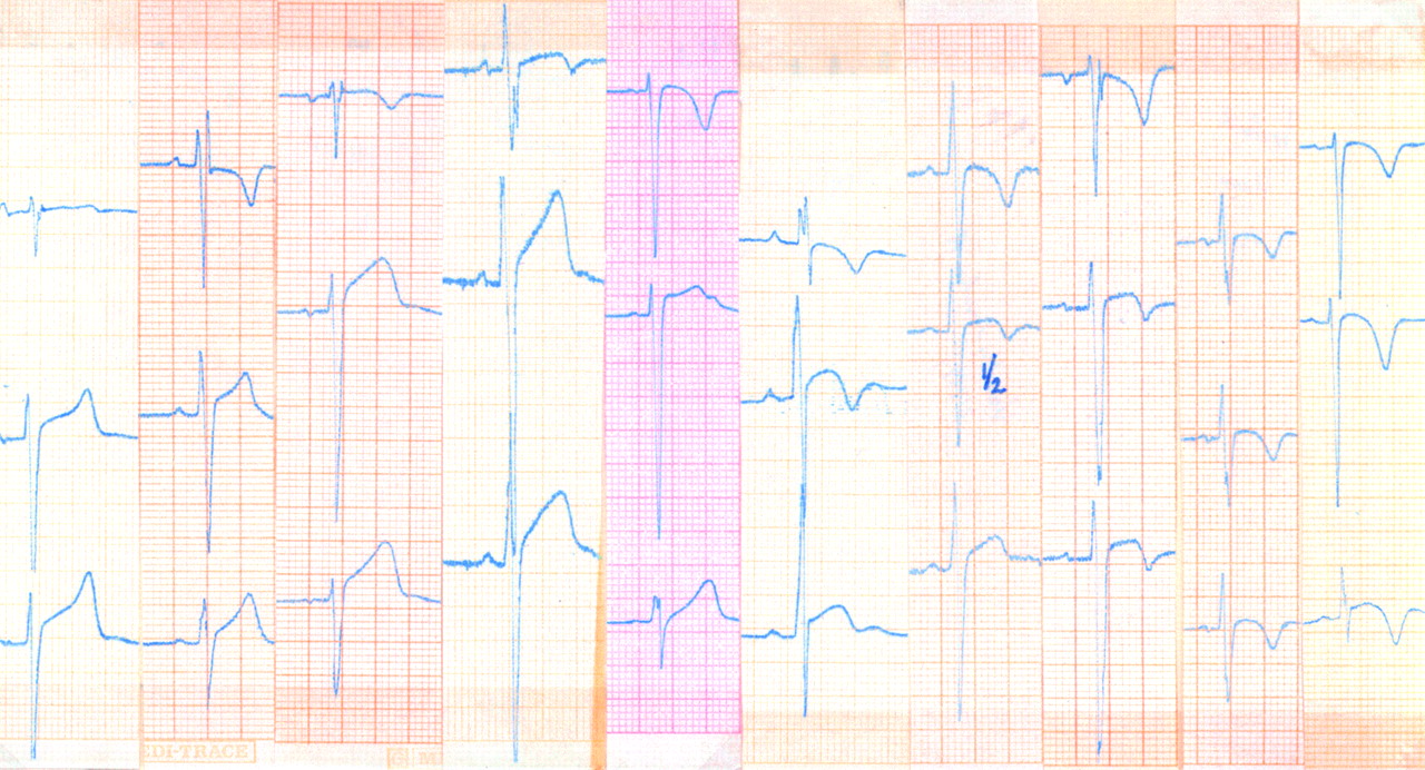
Figures 2 and 3 illustrate morphologic alterations in the right and left precordial leads, respectively.

Right precordial leads V1–3 in athletes. Note the variable QRS pattern in lead V1. The ST-T segments show variable combinations of ST elevation (with upward concavity to convexity) and upright, diphasic, or inverted T waves.

Left precordial leads V4–6 in athletes. Note the high R wave in some athletes and the variable pattern of the ST-T segment.
Ambulatory electrocardiography
The 12 lead ECG lasts only a few seconds, so that 24 hour ambulatory electrocardiography may give a better insight into the occurrence of rhythm and conduction alterations in athletes. Viitasalo and colleagues18 compared 35 highly trained male endurance athletes with 35 non-athletic matched controls. Heart rate was lower in athletes throughout the day and night. As shown in table 3 the lowest nocturnal heart rate ranged from 24–48 beats/min in the athletes and from 33–63 beats/min in the controls. Sinus pauses exceeding 2.0 s occurred in 37.1% and 5.7%, respectively, with the longest PP intervals of 2.76 s and 2.6 s. First degree atrioventricular block and second degree Möbitz type 1 block occurred more frequently in athletes. Atrioventricular dissociation and Möbitz type II block were not observed in controls but did occur in athletes. Disturbances were more prevalent during the night than during the day. The frequency of ventricular arrhythmias did not differ between athletes and controls.
Heart rate and frequency of cardiac events on ambulatory monitoring of athletes and non-athletes
Exercise electrocardiography
Dynamic exercise is associated with increased sympathetic drive and a reduction of vagal tone. Rhythm disturbances such as sinus bradycardia, sinus arrest, and wandering pacemaker, as well as the atrioventricular conduction defects, all disappear with exercise, frequently even on changing from the supine to the sitting or standing position. Repolarisation abnormalities as a rule do also normalise with exercise. These observations, together with the effects of pharmacological agents such as atropine, and other interventions such as the Valsalva manoeuvre, led to the conclusion that the described alterations are functional and not structural.
Clinical significance
To clarify the clinical significance of abnormal ECG patterns in athletes, Pelliccia and colleagues19 compared ECG findings with cardiac morphology assessed by echocardiography in 1005 consecutive athletes who were participating in 38 sporting disciplines. Based on a variety of criteria, ECG patterns were distinctly abnormal in 14%, mildly abnormal in 26%, and normal or with minor alterations in 60%. Abnormal ECGs were associated with male sex, younger age, endurance sports, and larger cardiac dimensions; structural cardiovascular diseases were rarely responsible for the abnormal ECG patterns in trained athletes. It was concluded that bizarre ECG patterns may be part of athlete’s heart syndrome. On the other hand, normal ECGs were highly predictive of an absence of cardiovascular abnormalities.
CONSIDERATIONS AND CONCERNS FOR THE FUTURE
Whereas athlete’s heart is at least partly caused by the training per se, twin studies revealed significant heritability of left ventricular wall thickness, so that cardiac alterations in athletes may be partly genetic.3 Some studies have found significant associations between left ventricular mass and genetic polymorphisms, such as in the renin angiotensin system. The possible genetic involvement in athlete’s heart should be further explored.
Newer non-invasive techniques will permit us to study myocardial metabolism in athletes. For example, Pluim and colleagues20 used magnetic resonance spectroscopy and concluded that strenuous training was not associated with pathological changes in myocardial high energy phosphate metabolism.
Finally, although athlete’s heart is usually considered to be physiologic, there is some concern that present day, high intensity training may contribute to the development of malignant ventricular arrhythmias and exercise related sudden death, but the possible role of ergogenic aids cannot be fully excluded. Also, the fact that the heart remains enlarged after the cessation of training in a number of athletes has raised concern.9
The ECG: key points
-
High intensity dynamic endurance sports are usually associated with electrocardiographic rhythm and conduction abnormalities, which result from the lower intrinsic heart rate and/or changes in parasympathetic and sympathetic tone
-
Structural cardiac adaptations induce morphological changes of the QRS complex
-
Repolarisation abnormalities result from structural changes and from parasympathetic predominance
-
Several ECG changes may mimic cardiac disease
Acknowledgments
The author gratefully acknowledges the secretarial assistance of N Ausseloos. R Fagard is holder of the Professor A Amery Chair in Hypertension Research.
REFERENCES
Linked Articles
- Miscellanea
- Miscellanea