Article Text

Abstract
Introduction Recent cadaveric [Van Sterenberg et al. 2011], histological [Spang et al. 2013], and clinical/surgical (Alfredson, 2011) studies lend support to the theory that the plantaris tendon may be involved in the aetiology and/or pathogenesis of midportion Achilles tendinopathy. Unfortunately, it is not easy to detect a plantaris tendon, especially when it is localised close to the medial side of the Achilles tendon.
Ultrasound+Doppler (US+CD) has been used for many years as a first line diagnostic tool to detect tendinosis-like changes in tendons [Ohberg et al. 2001, Fornage et al. 1986]. Although there is evidence that the plantaris tendon can be detected by ultrasound (US) [Mackay et al. 1990, Spina 2007], there are no studies comparing macroscopic findings with ultrasound findings. Recently, Ultrasound Tissue Characterisation (UTC), has been used to visualise Achilles tendon structure, and to quantify tendon matrix integrity [Van Schie et al. 2001].
The aim of this study was to compare US+CD and UTC findings with macroscopic surgical (in wound) findings in patients with chronic painful midportion
Method and Material In this study on 16 patients (11 men, mean age: 38 years and 5 women, mean age: 41 years) and 20 tendons (4 patients with bilateral tendons) with plantaris tendon involvement in mid-portion Achilles tendinopathy, we used Ultrasound and Colour Doppler (US+CD) and Ultrasound Tissue Characterisation (UTC) findings and compared these findings with the macroscopic findings (in wound).
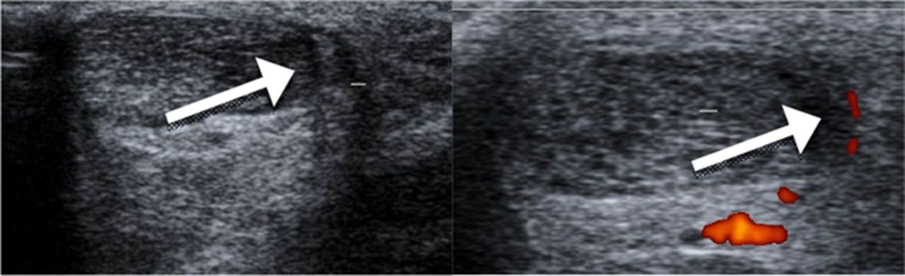
Results At surgery, in all 20 Achilles tendinopathy tendons, we found a thickened plantaris tendon located close to, or invaginating into, the medial side of the Achilles tendon. There was richly vascularised fat tissue in between the Achilles and plantaris tendons [fig 1]. For US+CD, 16/20 tendons had a tendon-like structure interpreted to be the plantaris tendon and localised high blood flow, in close relation to the medial side of the Achilles [fig 2]. For UTC, 19/20 tendons had disorganised (type 3 and 4) echopixels in the medial part of the Achilles tendon indicating possible plantaris tendon involvement [fig 3].
Conclusions US+CD can directly, and UTC indirectly, detect a plantaris tendon located close to the medial Achilles in a high percentage of patients with midportion Achilles tendinopathy.

Surgical findings

US/CD results: Plantaris tendon-like structure and high medial blood flow

UTC results: Focal disorganised echopixels in the medial part of midportion of Achilles
References Alfredson, Br J Sports Med. 2011;45:407–410
Alfredson, Br J Sports Med. 2011;45:1023–1025
Fornage, et al. Radiology. 1986;159:759–764
Mackay, et al. Br J Plast Surg. 1990;43:689–691
Ohberg, et al . Knee Surg Sports Traumatol Arthroosc 2001;9:233–288
Spina, J Can Chiropr Assoc. 2007;51:158–165
Van Sterkenburg, et al. J Anat. 2011;218:336–341
Van Schie, et al. Am J Vet Res. 2001;62:159–166