Article Text

Abstract
Introduction Platelet-Rich Plasma (PRP), an autologous derivative of whole blood that contains a supraphysiological concentration of platelets, has gained increasing attention [Alsousou 2009](1). An injection of platelets is thought to invoke an earlier and improved tissue healing response brought on by an increase in growth factors. The benefit of PRP has been supported by in vitro and animal studies in bone, cartilage, tendon and muscle [Engeberston, 2010] (2). To our knowledge there has been no research into the PRP effects in in vivo human Achilles. The aim of this study was to investigate the histological response of ruptured Achilles tendon treated with PRP.
Methods Tendon tissue biopsies were obtained from Achilles tendon rupture patients recruited in a clinical trial and in full compliance with HTA and Ethics Committee approval. Patients underwent tendon repair with/out PRP as part of the trial protocol. Needle biopsies were obtained under ultrasound guidance from the healing area of Achilles tendon 6 weeks post treatment. All samples were embedded in paraffin wax, sectioned and stained using H&E and Alcian Blue. Immunohistochemistry (IHC) markers were used to identify: collagen I and III, lymphocytes (CD45), proliferation (KI67) and blood vessels (CD34). Image analysis was performed using a light microscope (Zeiss AXIO Imager) equipped with a high resolution microscopy digital camera. Under 40x magnification, samples were systematically imaged to include the entire sample and avoid allocation bias. All images were blinded and IHC staining quantified using Image J software.
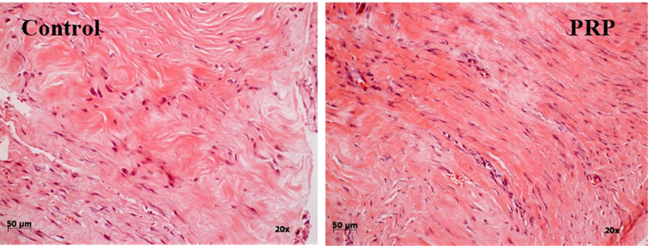
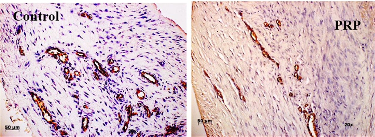
Results There was significantly higher cellularity and glycosaminoglycan content in PRP treated tendons (p = 0.01 and < 0.001 respectively) [Figures 1 and 2]. Fiber structure of the tissue was significantly better in the PRP group compared with control tissue (p < 0.001). Although both groups showed high collagen I staining, there was significantly higher content of collagen I in PRP when compared with control (p = 0.0079) while Collagen III content was not different (p = 1.0). The ratio of collagen III over collagen I was significantly lower in PRP (p = 0.007). There was no significant difference in CD45 expression (p = 0.33). However, PRP samples had fewer blood vessels than control (p = 0.023). The overall modified Bonar score of PRP samples was significantly lower than control [Table 1], which indicates improved early tendon healing.

H&E staining of control and PRP treated tendon tissue

CD34IHC stain examples

Modified Bonar score results
Conclusion To our knowledge this is the first study to report the histological and immunohistochemical response of human Achilles tendon to PRP therapy. The findings reveal that locally applied PRP in Achilles tendon rupture enhance the maturity of the healing tendon tissues by promoting better collagen I deposition, improved Collagen III/Collagen I ratio, decreased cellularity, less vascularity and higher glycosaminoglycan content when compared with control. These results may explain the early clinical improvement observed in these patients at week 6 onwards. Further work is required to determine the longer term effects of the use of PRP in musculoskeletal pathologies.
References 1 Alsousou et al. JBJS Br. 2009;91(8):987
2 Engebretsen et al. BJSM. 2010;44(15):1072